PELP1 Polyklonaler Antikörper
PELP1 Polyklonal Antikörper für IF, IHC, WB, ELISA
Wirt / Isotyp
Kaninchen / IgG
Getestete Reaktivität
human, Maus
Anwendung
WB, IHC, IF, ELISA
Konjugation
Unkonjugiert
Kat-Nr. : 30135-1-AP
Synonyme
Galerie der Validierungsdaten
Geprüfte Anwendungen
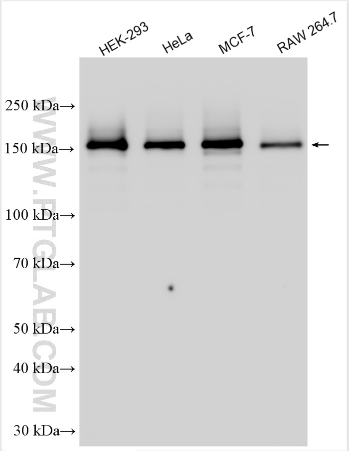
| Erfolgreiche Detektion in WB | HEK-293-Zellen, HeLa-Zellen, MCF-7-Zellen, RAW 264.7-Zellen |
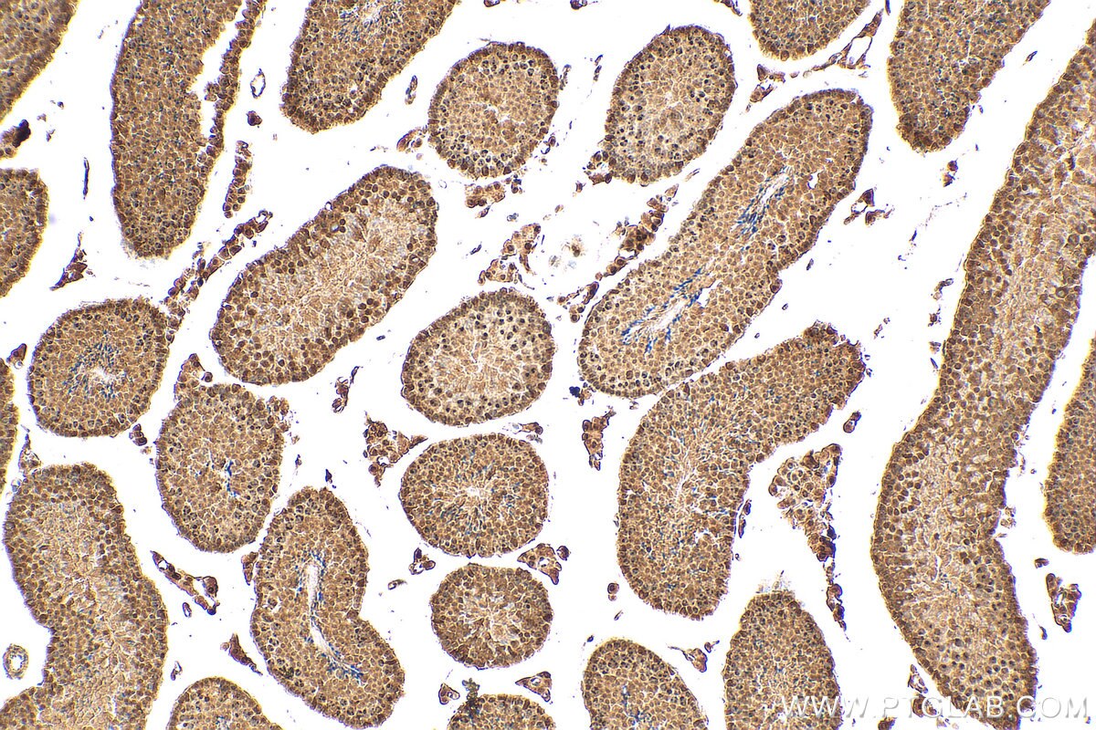
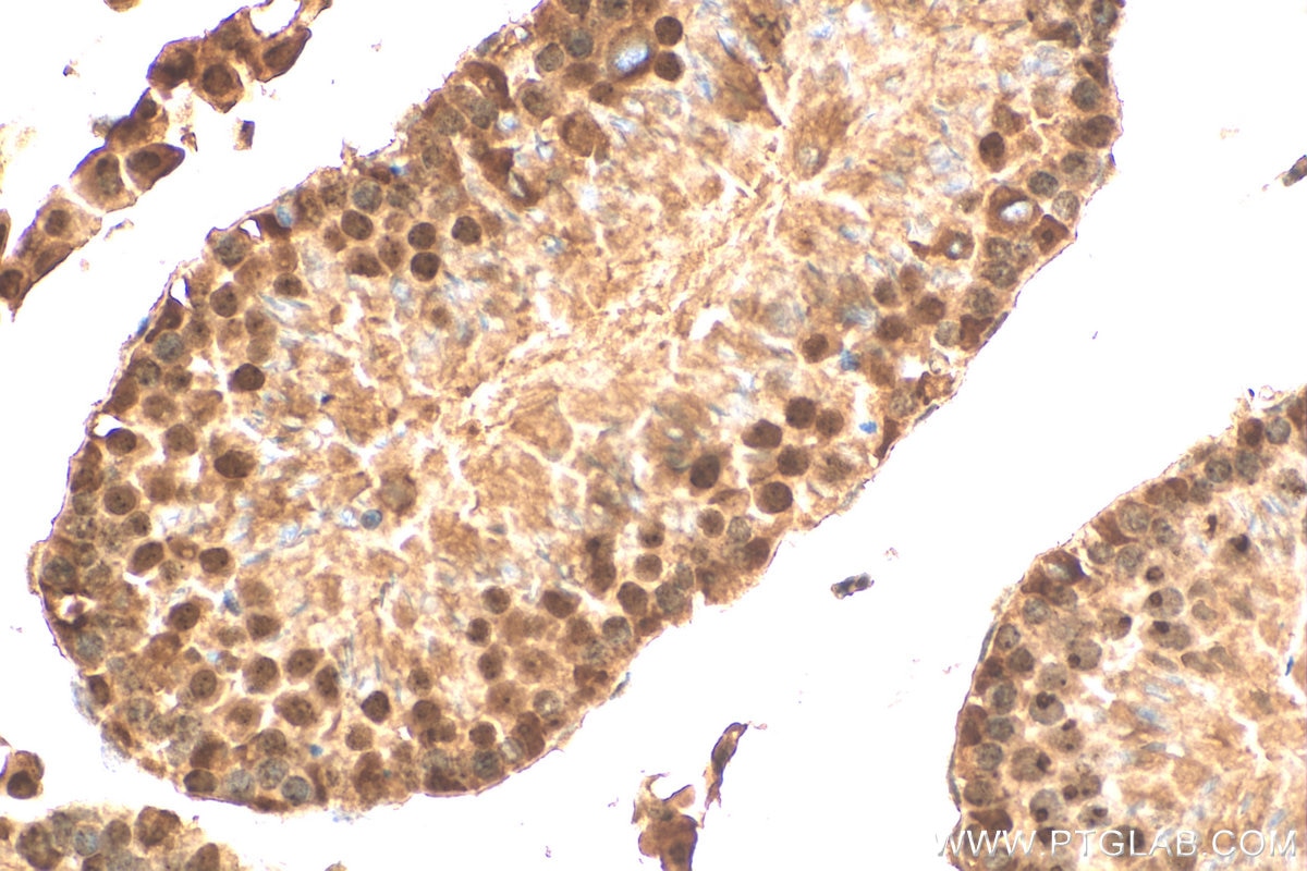
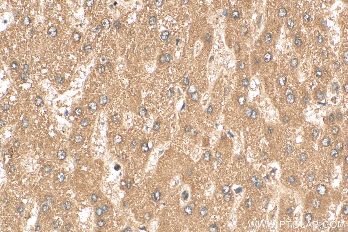
| Erfolgreiche Detektion in IHC | Maushodengewebe, humanes Lebergewebe Hinweis: Antigendemaskierung mit TE-Puffer pH 9,0 empfohlen. (*) Wahlweise kann die Antigendemaskierung auch mit Citratpuffer pH 6,0 erfolgen. |
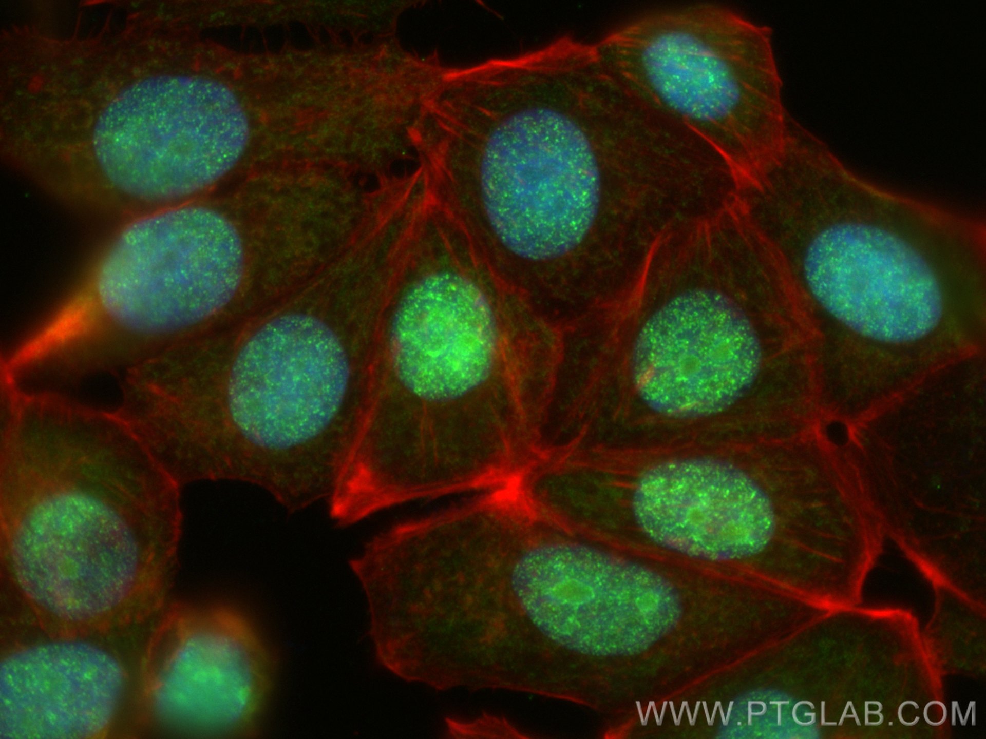
| Erfolgreiche Detektion in IF | MCF-7-Zellen |
Empfohlene Verdünnung
| Anwendung | Verdünnung |
|---|---|
| Western Blot (WB) | WB : 1:5000-1:50000 |
| Immunhistochemie (IHC) | IHC : 1:50-1:500 |
| Immunfluoreszenz (IF) | IF : 1:50-1:500 |
| It is recommended that this reagent should be titrated in each testing system to obtain optimal results. | |
| Sample-dependent, check data in validation data gallery | |
Produktinformation
30135-1-AP bindet in WB, IHC, IF, ELISA PELP1 und zeigt Reaktivität mit human, Maus
| Getestete Reaktivität | human, Maus |
| Wirt / Isotyp | Kaninchen / IgG |
| Klonalität | Polyklonal |
| Typ | Antikörper |
| Immunogen | PELP1 fusion protein Ag32720 |
| Vollständiger Name | proline, glutamate and leucine rich protein 1 |
| Berechnetes Molekulargewicht | 1130 aa, 120 kDa |
| Beobachtetes Molekulargewicht | 160 kDa |
| GenBank-Zugangsnummer | BC069058 |
| Gene symbol | PELP1 |
| Gene ID (NCBI) | 27043 |
| Konjugation | Unkonjugiert |
| Form | Liquid |
| Reinigungsmethode | Antigen-Affinitätsreinigung |
| Lagerungspuffer | PBS mit 0.02% Natriumazid und 50% Glycerin pH 7.3. |
| Lagerungsbedingungen | Bei -20°C lagern. Nach dem Versand ein Jahr lang stabil Aliquotieren ist bei -20oC Lagerung nicht notwendig. 20ul Größen enthalten 0,1% BSA. |
Hintergrundinformationen
PELP1 was first identified as a 160 kDa protein in a screen for Src homology 2 (SH2) domain-binding proteins. PELP1 is overexpressed in 60-80% of breast tumors and plays important roles in both ER genomic and non-genomic signaling. In vivo, PELP1 subcellular localization is primarily nuclear in normal breast tissue, but it is localized to the cytoplasm in about 40% of invasive breast tumors. In the nucleus, PELP1 interacts with a number of transcription factors. The proto-oncogenic functions of PELP1 involve different cellular processes including epigenetic modifications leading to ER transactivation and breast cancer progression. Furthermore, PELP1 activates kinase cascades in the cytoplasm such as MAPK activation via c-Src and PI3K signaling.
Protokolle
| Produktspezifische Protokolle | |
|---|---|
| WB protocol for PELP1 antibody 30135-1-AP | Protokoll herunterladen |
| IHC protocol for PELP1 antibody 30135-1-AP | Protokoll herunterladen |
| IF protocol for PELP1 antibody 30135-1-AP | Protokoll herunterladen |
| Standard-Protokolle | |
|---|---|
| Klicken Sie hier, um unsere Standardprotokolle anzuzeigen |
