PD-1/CD279 Monoklonaler Antikörper
PD-1/CD279 Monoklonal Antikörper für FC, IF
Wirt / Isotyp
Maus / IgG2b
Getestete Reaktivität
human, Maus, Ratte
Anwendung
IF, FC
Konjugation
CoraLite® Plus 488 Fluorescent Dye
CloneNo.
4H4D1
Kat-Nr. : CL488-66220
Synonyme
Galerie der Validierungsdaten
Geprüfte Anwendungen
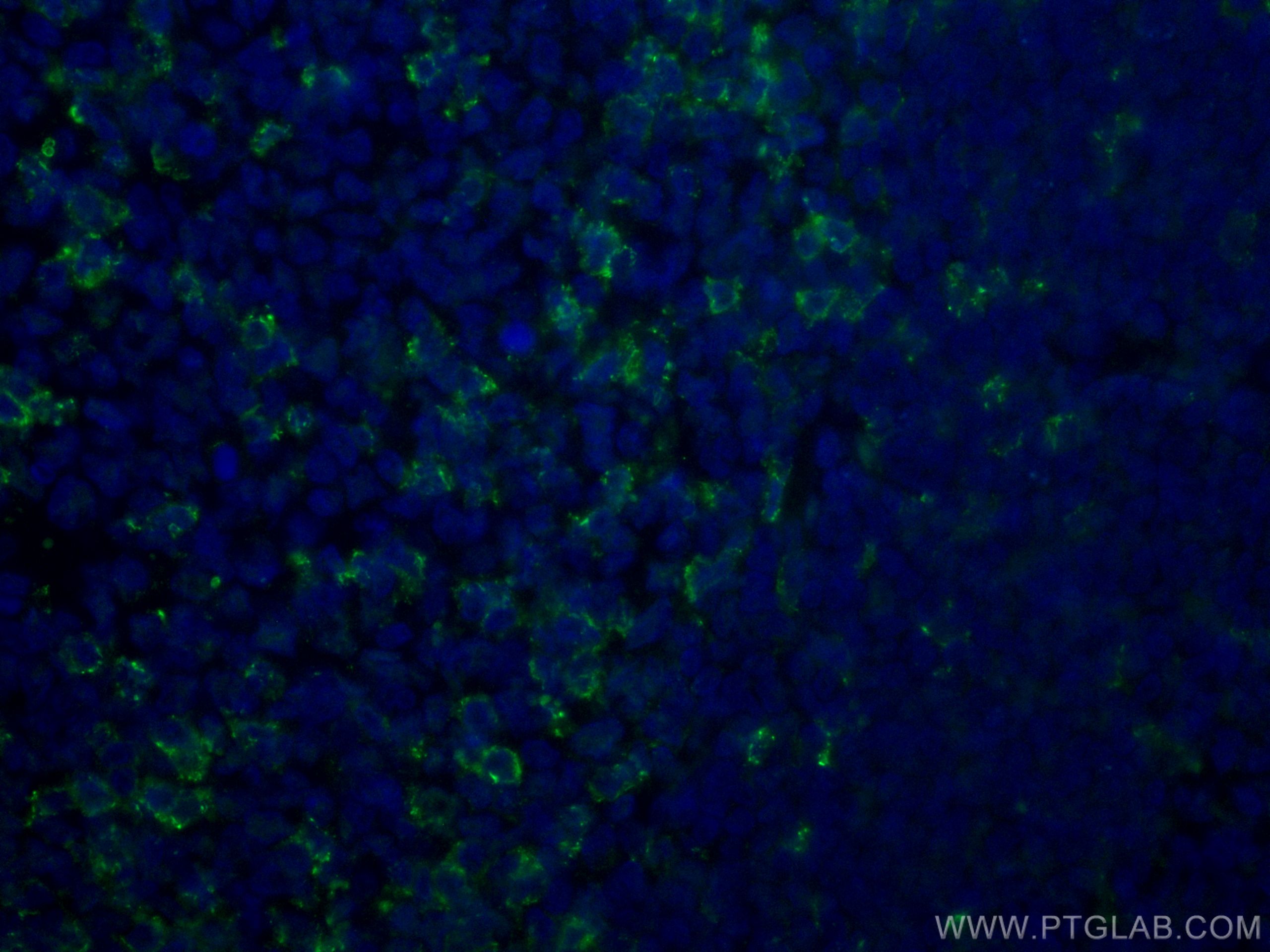
| Erfolgreiche Detektion in IF | humanes Tonsillitisgewebe |
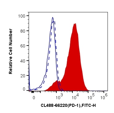
| Erfolgreiche Detektion in FC | CD3 antibody treated Jurkat cells |
Empfohlene Verdünnung
| Anwendung | Verdünnung |
|---|---|
| Immunfluoreszenz (IF) | IF : 1:50-1:500 |
| Durchflusszytometrie (FC) | FC : 0.40 ug per 10^6 cells in a 100 µl suspension |
| It is recommended that this reagent should be titrated in each testing system to obtain optimal results. | |
| Sample-dependent, check data in validation data gallery | |
Produktinformation
CL488-66220 bindet in IF, FC PD-1/CD279 und zeigt Reaktivität mit human, Maus, Ratten
| Getestete Reaktivität | human, Maus, Ratte |
| Wirt / Isotyp | Maus / IgG2b |
| Klonalität | Monoklonal |
| Typ | Antikörper |
| Immunogen | PD-1/CD279 fusion protein Ag12470 |
| Vollständiger Name | programmed cell death 1 |
| Berechnetes Molekulargewicht | 288 aa, 32 kDa |
| GenBank-Zugangsnummer | BC074740 |
| Gene symbol | PDCD1 |
| Gene ID (NCBI) | 5133 |
| Konjugation | CoraLite® Plus 488 Fluorescent Dye |
| Excitation/Emission maxima wavelengths | 493 nm / 522 nm |
| Form | Liquid |
| Reinigungsmethode | Protein-A-Reinigung |
| Lagerungspuffer | BS mit 50% Glyzerin, 0,05% Proclin300, 0,5% BSA, pH 7,3. |
| Lagerungsbedingungen | Bei -20°C lagern. Vor Licht schützen. Nach dem Versand ein Jahr stabil. Aliquotieren ist bei -20oC Lagerung nicht notwendig. 20ul Größen enthalten 0,1% BSA. |
Hintergrundinformationen
Programmed cell death 1 (PD-1, also known as CD279) is an immunoinhibitory receptor that belongs to the CD28/CTLA-4 subfamily of the Ig superfamily. It is a 288 amino acid (aa) type I transmembrane protein composed of one Ig superfamily domain, a stalk, a transmembrane domain, and an intracellular domain containing an immunoreceptor tyrosine-based inhibitory motif (ITIM) as well as an immunoreceptor tyrosine-based switch motif (ITSM) (PMID: 18173375). PD-1 is expressed during thymic development and is induced in a variety of hematopoietic cells in the periphery by antigen receptor signaling and cytokines (PMID: 20636820). Engagement of PD-1 by its ligands PD-L1 or PD-L2 transduces a signal that inhibits T-cell proliferation, cytokine production, and cytolytic function (PMID: 19426218). It is critical for the regulation of T cell function during immunity and tolerance. Blockade of PD-1 can overcome immune resistance and also has been shown to have antitumor activity (PMID: 22658127; 23169436). The calculated molecular weight of PD-1 is 32 kDa. It has been reported that PD-1 is heavily glycosylated and migrates with an apparent molecular mass of 47-55 kDa on SDS-PAGE (PMID: 8671665; 17640856; 17003438). This anitbody is CL488(Ex/Em 488 nm/515 nm) conjugated.
Protokolle
| Produktspezifische Protokolle | |
|---|---|
| IF protocol for CL Plus 488 PD-1/CD279 antibody CL488-66220 | Protokoll herunterladen |
| Standard-Protokolle | |
|---|---|
| Klicken Sie hier, um unsere Standardprotokolle anzuzeigen |


