OPA3 Monoklonaler Antikörper
OPA3 Monoklonal Antikörper für WB, ELISA
Wirt / Isotyp
Maus / IgG1
Getestete Reaktivität
human
Anwendung
WB, ELISA
Konjugation
Unkonjugiert
CloneNo.
5A3F9
Kat-Nr. : 68241-1-Ig
Synonyme
Galerie der Validierungsdaten
Geprüfte Anwendungen
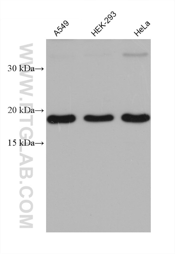
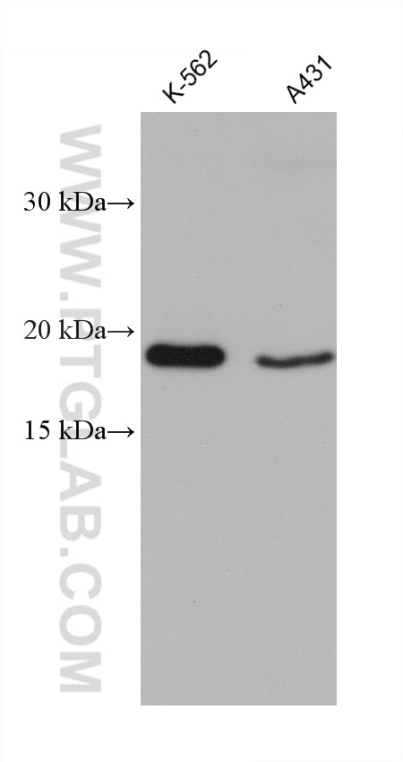
| Erfolgreiche Detektion in WB | A549-Zellen, A431-Zellen, HEK-293-Zellen, HeLa-Zellen, K-562-Zellen |
Empfohlene Verdünnung
| Anwendung | Verdünnung |
|---|---|
| Western Blot (WB) | WB : 1:1000-1:6000 |
| It is recommended that this reagent should be titrated in each testing system to obtain optimal results. | |
| Sample-dependent, check data in validation data gallery | |
Produktinformation
68241-1-Ig bindet in WB, ELISA OPA3 und zeigt Reaktivität mit human
| Getestete Reaktivität | human |
| Wirt / Isotyp | Maus / IgG1 |
| Klonalität | Monoklonal |
| Typ | Antikörper |
| Immunogen | OPA3 fusion protein Ag8173 |
| Vollständiger Name | optic atrophy 3 (autosomal recessive, with chorea and spastic paraplegia) |
| Berechnetes Molekulargewicht | 179 aa, 20 kDa |
| Beobachtetes Molekulargewicht | 20 kDa |
| GenBank-Zugangsnummer | BC005059 |
| Gene symbol | OPA3 |
| Gene ID (NCBI) | 80207 |
| Konjugation | Unkonjugiert |
| Form | Liquid |
| Reinigungsmethode | Protein-G-Reinigung |
| Lagerungspuffer | PBS mit 0.02% Natriumazid und 50% Glycerin pH 7.3. |
| Lagerungsbedingungen | Bei -20°C lagern. Nach dem Versand ein Jahr lang stabil Aliquotieren ist bei -20oC Lagerung nicht notwendig. 20ul Größen enthalten 0,1% BSA. |
Hintergrundinformationen
The OPA3 cDNA encodes a deduced 179-amino acid protein. Northern blot analysis demonstrated a primary transcript of approximately 5.0 kb that was ubiquitously expressed, most prominently in skeletal muscle and kidney. Mutations in this gene have been shown to result in 3-methylglutaconic aciduria type III and autosomal dominant optic atrophy and cataract.
Protokolle
| Produktspezifische Protokolle | |
|---|---|
| WB protocol for OPA3 antibody 68241-1-Ig | Protokoll herunterladen |
| Standard-Protokolle | |
|---|---|
| Klicken Sie hier, um unsere Standardprotokolle anzuzeigen |


