- Featured Product
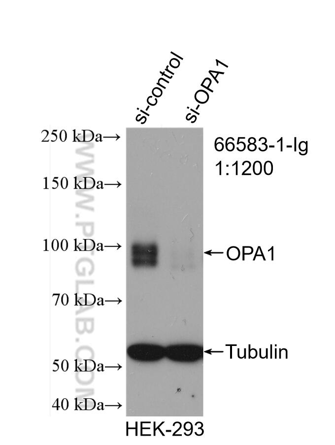
- KD/KO Validated
OPA1 Monoklonaler Antikörper
OPA1 Monoklonal Antikörper für IHC, WB, ELISA
Wirt / Isotyp
Maus / IgG2b
Getestete Reaktivität
Hausschwein, human, Maus, Ratte und mehr (1)
Anwendung
WB, IHC, IF, ELISA
Konjugation
Unkonjugiert
CloneNo.
1B2D8
Kat-Nr. : 66583-1-Ig
Synonyme
Galerie der Validierungsdaten
Geprüfte Anwendungen
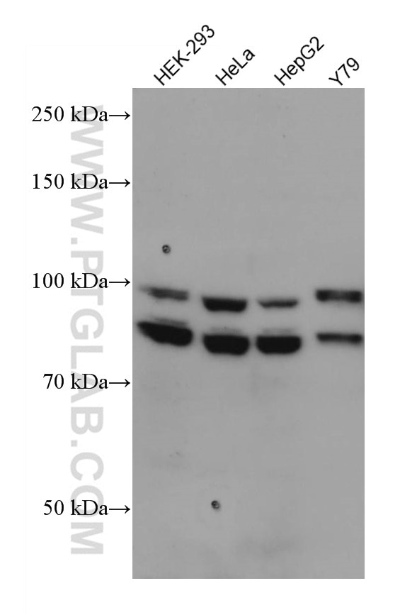
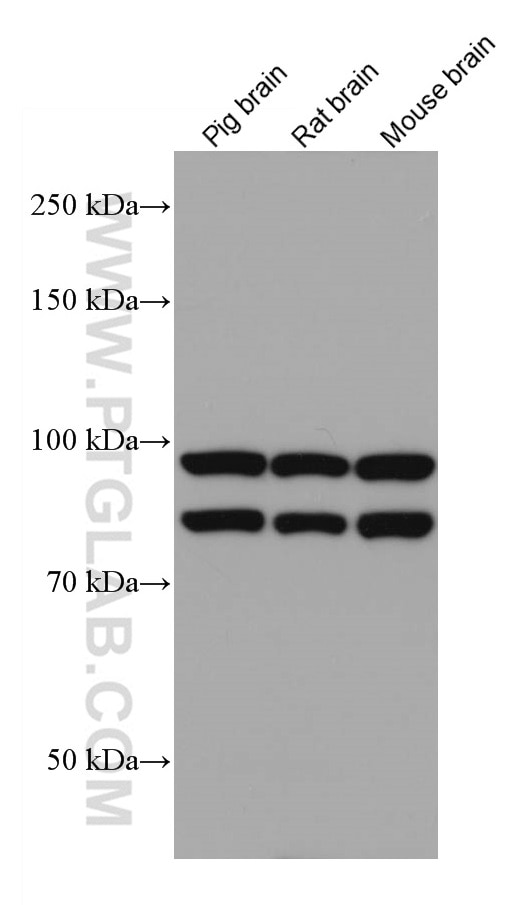
| Erfolgreiche Detektion in WB | HEK-293-Zellen, Hausschwein-Hirngewebe, HeLa-Zellen, HepG2-Zellen, Maushirngewebe, Rattenhirngewebe, Y79-Zellen |
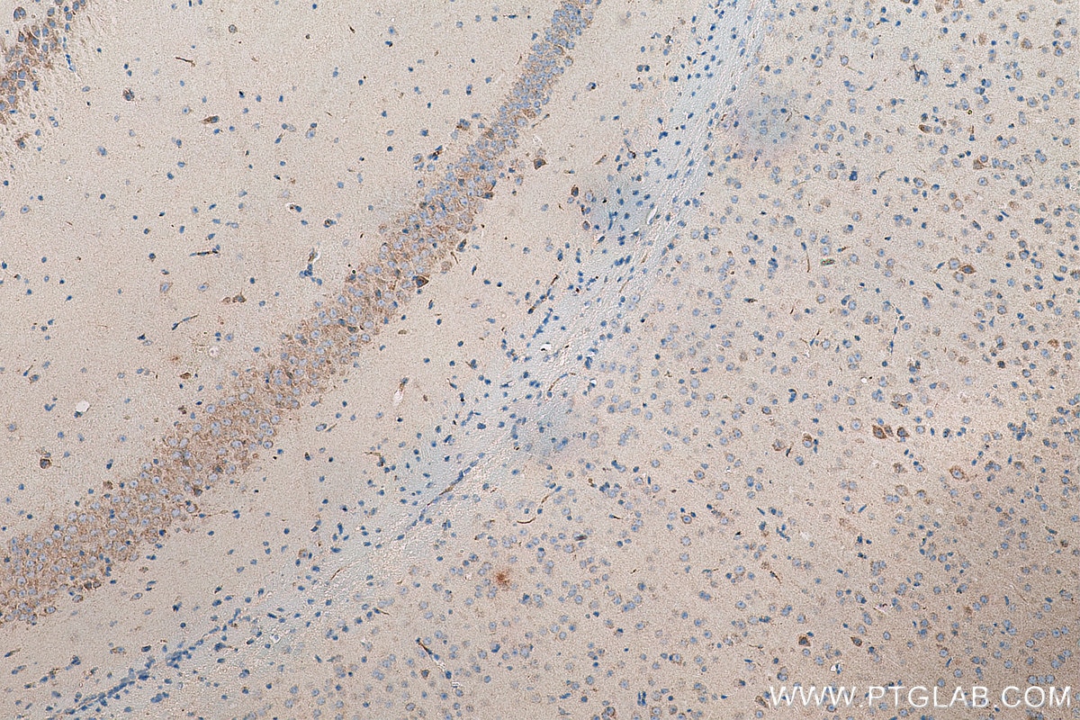
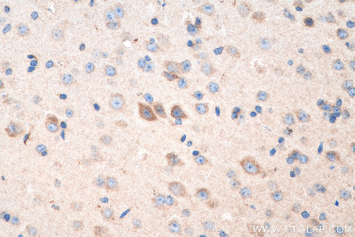
| Erfolgreiche Detektion in IHC | Maushirngewebe Hinweis: Antigendemaskierung mit TE-Puffer pH 9,0 empfohlen. (*) Wahlweise kann die Antigendemaskierung auch mit Citratpuffer pH 6,0 erfolgen. |
Empfohlene Verdünnung
| Anwendung | Verdünnung |
|---|---|
| Western Blot (WB) | WB : 1:500-1:2000 |
| Immunhistochemie (IHC) | IHC : 1:400-1:1600 |
| It is recommended that this reagent should be titrated in each testing system to obtain optimal results. | |
| Sample-dependent, check data in validation data gallery | |
Veröffentlichte Anwendungen
| KD/KO | See 1 publications below |
| WB | See 13 publications below |
| IF | See 1 publications below |
Produktinformation
66583-1-Ig bindet in WB, IHC, IF, ELISA OPA1 und zeigt Reaktivität mit Hausschwein, human, Maus, Ratten
| Getestete Reaktivität | Hausschwein, human, Maus, Ratte |
| In Publikationen genannte Reaktivität | human, Fisch, Maus, Ratte |
| Wirt / Isotyp | Maus / IgG2b |
| Klonalität | Monoklonal |
| Typ | Antikörper |
| Immunogen | OPA1 fusion protein Ag26868 |
| Vollständiger Name | optic atrophy 1 (autosomal dominant) |
| Berechnetes Molekulargewicht | 960 aa, 112 kDa |
| Beobachtetes Molekulargewicht | 100 kDa and 80-90 kDa |
| GenBank-Zugangsnummer | BC075805 |
| Gene symbol | OPA1 |
| Gene ID (NCBI) | 4976 |
| Konjugation | Unkonjugiert |
| Form | Liquid |
| Reinigungsmethode | Protein-A-Reinigung |
| Lagerungspuffer | PBS mit 0.02% Natriumazid und 50% Glycerin pH 7.3. |
| Lagerungsbedingungen | Bei -20°C lagern. Nach dem Versand ein Jahr lang stabil Aliquotieren ist bei -20oC Lagerung nicht notwendig. 20ul Größen enthalten 0,1% BSA. |
Hintergrundinformationen
OPA1 is a nuclear-encoded mitochondrial protein with similarity to dynamin-related GTPases. OPA1 localizes to the inner mitochondrial membrane and helps regulate mitochondrial stability and energy output. This protein also sequesters cytochrome c. OPA1 is associated with the inner membrane and protects cells from apoptosis by regulating inner membrane dynamics. Mutation of OPA1 causes the disease dominant optic atrophy, a degeneration of the retinal ganglion cells. OPA1 undergoes complex posttranscriptional regulation and posttranslational proteolysis. OPA1 is regulated by proteolytic cleavage, which degrades long OPA1 isoforms into short isoforms. The gene OPA1 can be cleaved into some chains with MW 100 kDa and 80-90 kDa.
Protokolle
| Produktspezifische Protokolle | |
|---|---|
| WB protocol for OPA1 antibody 66583-1-Ig | Protokoll herunterladen |
| IHC protocol for OPA1 antibody 66583-1-Ig | Protokoll herunterladen |
| Standard-Protokolle | |
|---|---|
| Klicken Sie hier, um unsere Standardprotokolle anzuzeigen |
Publikationen
| Species | Application | Title |
|---|---|---|
J Physiol Sci Cardioprotective responses to aerobic exercise-induced physiological hypertrophy in zebrafish heart | ||
Behav Brain Res Protective effect of exercise training against the progression of Alzheimer's disease in 3xTg-AD mice. | ||
Eur J Pharmacol Dihydromyricetin contributes to weight loss via pro-browning mediated by mitochondrial fission in white adipose | ||
Cell Death Dis Cadmium-induced apoptosis of Leydig cells is mediated by excessive mitochondrial fission and inhibition of mitophagy | ||
Aging (Albany NY) Jian-Pi-Yi-Shen decoction inhibits mitochondria-dependent granulosa cell apoptosis in a rat model of POF |
