MYO6 Polyklonaler Antikörper
MYO6 Polyklonal Antikörper für IF, IHC, IP, WB,ELISA
Wirt / Isotyp
Kaninchen / IgG
Getestete Reaktivität
human, Maus
Anwendung
WB, IP, IHC, IF, ELISA
Konjugation
Unkonjugiert
Kat-Nr. : 26778-1-AP
Synonyme
Galerie der Validierungsdaten
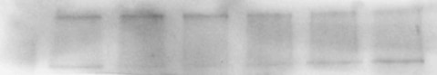
 at dilution of 1:500 incubated at room temperature for 1.5 hours.")
 with PC-3 cells lysate 2000 ug.")
 at dilution of 1:200 (under 40x lens). Heat mediated antigen retrieval with Tris-EDTA buffer (pH 9.0).")
 at dilution of 1:200 (under 40x lens).")
 fixed mouse small intestine tissue using 26778-1-AP (MYO6 antibody) at dilution of 1:50 and Alexa Fluor 488-Conjugated AffiniPure Goat Anti-Rabbit IgG(H+L).")
Geprüfte Anwendungen
| Erfolgreiche Detektion in WB | Maus-Dünndarmgewebe |
| Erfolgreiche IP | PC-3-Zellen |
| Erfolgreiche Detektion in IHC | humanes Prostatakarzinomgewebe, humanes Dünndarmgewebe Hinweis: Antigendemaskierung mit TE-Puffer pH 9,0 empfohlen. (*) Wahlweise kann die Antigendemaskierung auch mit Citratpuffer pH 6,0 erfolgen. |
| Erfolgreiche Detektion in IF | Maus-Dünndarmgewebe |
Empfohlene Verdünnung
| Anwendung | Verdünnung |
|---|---|
| Western Blot (WB) | WB : 1:500-1:1000 |
| Immunpräzipitation (IP) | IP : 0.5-4.0 ug for 1.0-3.0 mg of total protein lysate |
| Immunhistochemie (IHC) | IHC : 1:50-1:500 |
| Immunfluoreszenz (IF) | IF : 1:50-1:500 |
| It is recommended that this reagent should be titrated in each testing system to obtain optimal results. | |
| Sample-dependent, check data in validation data gallery | |
Veröffentlichte Anwendungen
| KD/KO | See 1 publications below |
| WB | See 3 publications below |
| IHC | See 2 publications below |
| IF | See 2 publications below |
Produktinformation
26778-1-AP bindet in WB, IP, IHC, IF, ELISA MYO6 und zeigt Reaktivität mit human, Maus
| Getestete Reaktivität | human, Maus |
| In Publikationen genannte Reaktivität | human, Maus |
| Wirt / Isotyp | Kaninchen / IgG |
| Klonalität | Polyklonal |
| Typ | Antikörper |
| Immunogen | MYO6 fusion protein Ag24906 |
| Vollständiger Name | myosin VI |
| Berechnetes Molekulargewicht | 1285 aa, 149 kDa |
| Beobachtetes Molekulargewicht | 145-150 kDa |
| GenBank-Zugangsnummer | BC146764 |
| Gene symbol | MYO6 |
| Gene ID (NCBI) | 4646 |
| Konjugation | Unkonjugiert |
| Form | Liquid |
| Reinigungsmethode | Antigen-Affinitätsreinigung |
| Lagerungspuffer | PBS mit 0.02% Natriumazid und 50% Glycerin pH 7.3. |
| Lagerungsbedingungen | Bei -20°C lagern. Nach dem Versand ein Jahr lang stabil Aliquotieren ist bei -20oC Lagerung nicht notwendig. 20ul Größen enthalten 0,1% BSA. |
Hintergrundinformationen
MYO6, an actin-based motor protein, is the only myosin known to move toward the minus end of actin filaments. MYO6 is highly expressed in the inner and outer hair cells of the ear, retina, and polarized epithelial cells such as kidney proximal tubule cells and intestinal enterocytes. And it participates in a wide range of biological processes within cells, including clathrin-mediated endocytosis, vesicular membrane traffic, polarized secretion, and autophagy (PMID: 23620821; PMID: 28591580). Previous studies showed that MYO6 is upregulated in various types of cancer, and it has been widely reported to contribute to tumor cell migration and metastasis. Some articles indicate that MYO6 is associated with prostate cancer, lung cancer, human colorectal cancer and gastric cancer (PMID: 29022908).
Protokolle
| Produktspezifische Protokolle | |
|---|---|
| WB protocol for MYO6 antibody 26778-1-AP | Protokoll herunterladen |
| IHC protocol for MYO6 antibody 26778-1-AP | Protokoll herunterladen |
| IF protocol for MYO6 antibody 26778-1-AP | Protokoll herunterladen |
| IP protocol for MYO6 antibody 26778-1-AP | Protokoll herunterladen |
| Standard-Protokolle | |
|---|---|
| Klicken Sie hier, um unsere Standardprotokolle anzuzeigen |
Publikationen
| Species | Application | Title |
|---|---|---|
Nat Commun Structure of Myosin VI/Tom1 complex reveals a cargo recognition mode of Myosin VI for tethering. | ||
Cell Signal Elevated expression of myosin VI contributes to breast cancer progression via MAPK/ERK signaling pathway
| ||
Nat Commun MYL3 protects chondrocytes from senescence by inhibiting clathrin-mediated endocytosis and activating of Notch signaling | ||
Int J Toxicol Copper Chaperone Atox1 Protected the Cochlea From Cisplatin by Regulating the Copper Transport Family and Cell Cycle |
Rezensionen
The reviews below have been submitted by verified Proteintech customers who received an incentive forproviding their feedback.
FH Wojciech (Verified Customer) (04-20-2023) | We used this antibody to assess protein levels in human T cells. We got a good signal with 1:1000 dilution with observed molecular weight at ~150kDa.
|
FH Sara (Verified Customer) (10-25-2020) | B cells (mouse) in RIPA buffer. 10 µg of protein per lane were loaded on a 10% PAGE-SDS gel. Transfer is not optimised for high MW proteins, so I could get a better blot. Two bands are detected, one of 150 kDa and one > 200 kDa.
 |

