MAP2 Polyklonaler Antikörper
MAP2 Polyklonal Antikörper für IF
Wirt / Isotyp
Kaninchen / IgG
Getestete Reaktivität
human, Maus, Ratte
Anwendung
IF
Konjugation
CoraLite® Plus 488 Fluorescent Dye
Kat-Nr. : CL488-17490
Synonyme
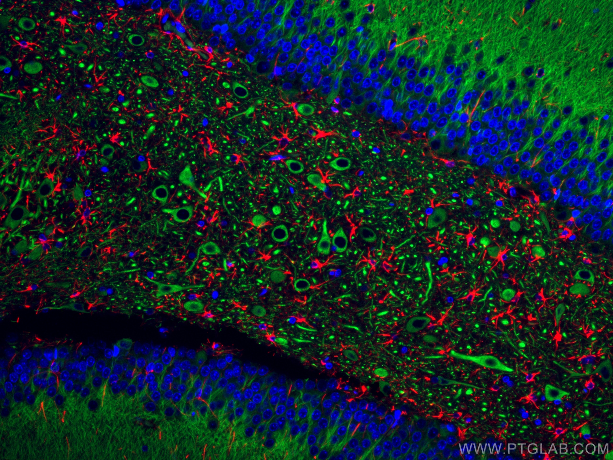
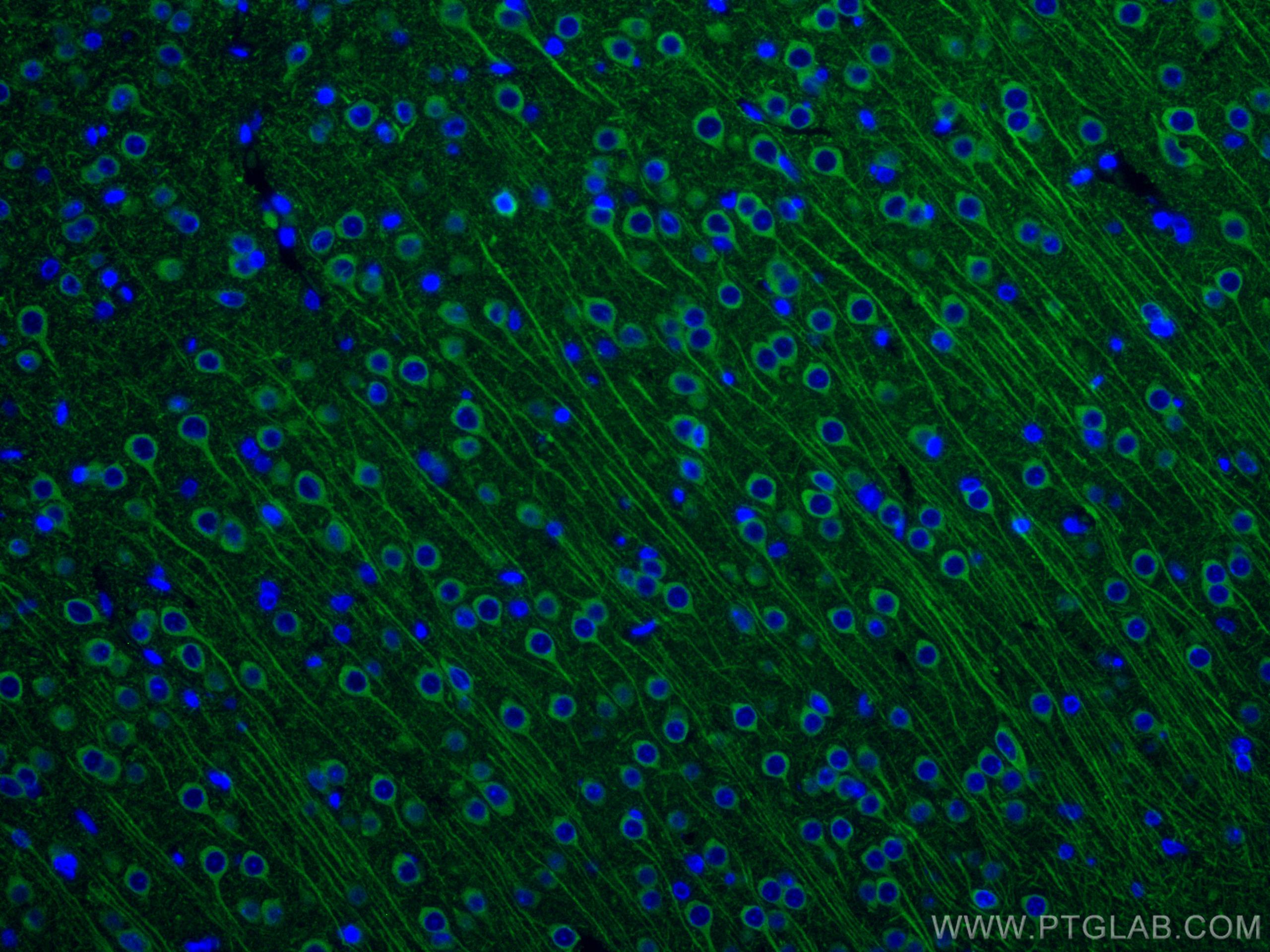
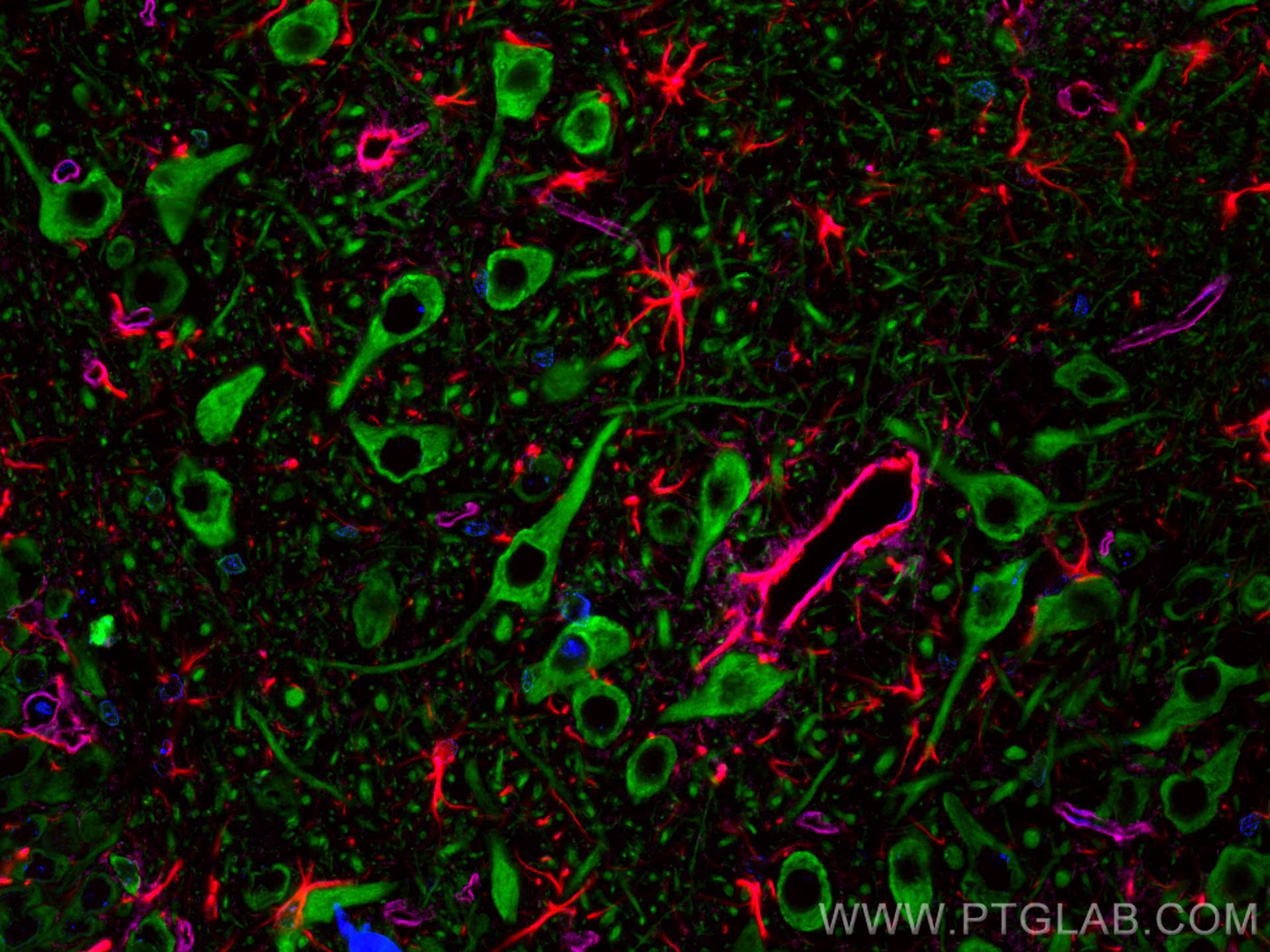
Galerie der Validierungsdaten
Geprüfte Anwendungen
| Erfolgreiche Detektion in IF | Rattenhirngewebe |
Empfohlene Verdünnung
| Anwendung | Verdünnung |
|---|---|
| Immunfluoreszenz (IF) | IF : 1:50-1:500 |
| It is recommended that this reagent should be titrated in each testing system to obtain optimal results. | |
| Sample-dependent, check data in validation data gallery | |
Produktinformation
CL488-17490 bindet in IF MAP2 und zeigt Reaktivität mit human, Maus, Ratten
| Getestete Reaktivität | human, Maus, Ratte |
| In Publikationen genannte Reaktivität | Maus |
| Wirt / Isotyp | Kaninchen / IgG |
| Klonalität | Polyklonal |
| Typ | Antikörper |
| Immunogen | MAP2 fusion protein Ag11580 |
| Vollständiger Name | microtubule-associated protein 2 |
| Berechnetes Molekulargewicht | 200 kDa |
| Beobachtetes Molekulargewicht | |
| GenBank-Zugangsnummer | BC038857 |
| Gene symbol | MAP2 |
| Gene ID (NCBI) | 4133 |
| Konjugation | CoraLite® Plus 488 Fluorescent Dye |
| Excitation/Emission maxima wavelengths | 493 nm / 522 nm |
| Form | Liquid |
| Reinigungsmethode | Antigen-Affinitätsreinigung |
| Lagerungspuffer | BS mit 50% Glyzerin, 0,05% Proclin300, 0,5% BSA, pH 7,3. |
| Lagerungsbedingungen | Bei -20°C lagern. Vor Licht schützen. Nach dem Versand ein Jahr stabil. Aliquotieren ist bei -20oC Lagerung nicht notwendig. 20ul Größen enthalten 0,1% BSA. |
Hintergrundinformationen
Microtubule-associated protein 2 (MAP2) is a tubulin binding protein regulating the spacing and stability of microtubules and contributing to elongation of dendrites.
What is the molecular weight of MAP2? Is MAP2 post-translationally modified?
MAP2 has multiple isoforms that arise from alternative splicing (PMID: 3121794, 7854050, and 10383434). They are classified into two groups - MAP2A and MAP2B, which are known as high molecular weight (HMW) isoforms, run as ~280 kDa species, while low molecular weight (LMW) isoforms MAP2C and MAP2D are around ~70 kDa. MAP2 proteins are heavily phosphorylated, which contributes to a large discrepancy between their predicted and observed molecular weight in SDS-PAGE (220 vs 280 kDa for HMW forms).
What is the tissue expression pattern of MAP2? What is the subcellular localization of MAP2?
MAP2 isoforms differ in their tissue and developmental expression pattern (PMID: 2469170 and 3898077). In the brain, MAP2B is widely expressed during and post development, MAP2A is expressed postnatally, while MAP2C is present only in the early development except of present in photosensitive cells of the adult retina and in the olfactory system. MAP proteins are highly expressed in the CNS found in cell bodies and dendrites of neurons, in dorsal root ganglion, reactive glia, and in the testis (PMID: 9588626). In neurons, MAP2 proteins are found in the cell body and dendrites, where they associate with microtubules, while they can also be present in the nuclei of testicular cells.
Can MAP2 be used as a neuronal marker?
MAP2 proteins are abundantly expressed in neurons. MAP2 is frequently used as a dendritic marker because it is present in the cell body and dendrites of neurons but absent in axons (PMID: 28413822).
Protokolle
| Produktspezifische Protokolle | |
|---|---|
| IF protocol for CL Plus 488 MAP2 antibody CL488-17490 | Protokoll herunterladen |
| Standard-Protokolle | |
|---|---|
| Klicken Sie hier, um unsere Standardprotokolle anzuzeigen |
Publikationen
| Species | Application | Title |
|---|---|---|
Antioxidants (Basel) Cellular Localization of Kynurenine 3-Monooxygenase in the Brain: Challenging the Dogma. |


