- Featured Product
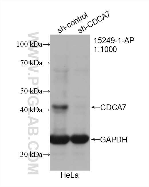
- KD/KO Validated
CDCA7 Polyklonaler Antikörper
CDCA7 Polyklonal Antikörper für IF, IP, WB, ELISA
Wirt / Isotyp
Kaninchen / IgG
Getestete Reaktivität
human, Maus, Ratte
Anwendung
WB, IP, IHC, IF, ELISA
Konjugation
Unkonjugiert
Kat-Nr. : 15249-1-AP
Synonyme
Galerie der Validierungsdaten
Geprüfte Anwendungen
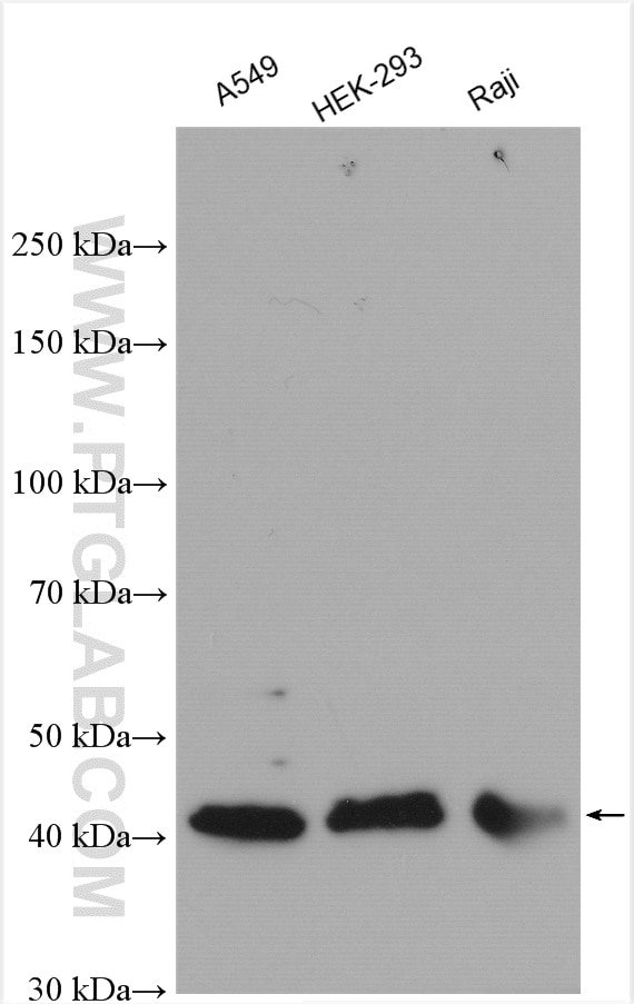
| Erfolgreiche Detektion in WB | A549-Zellen, HEK-293-Zellen, HeLa-Zellen, Raji-Zellen |
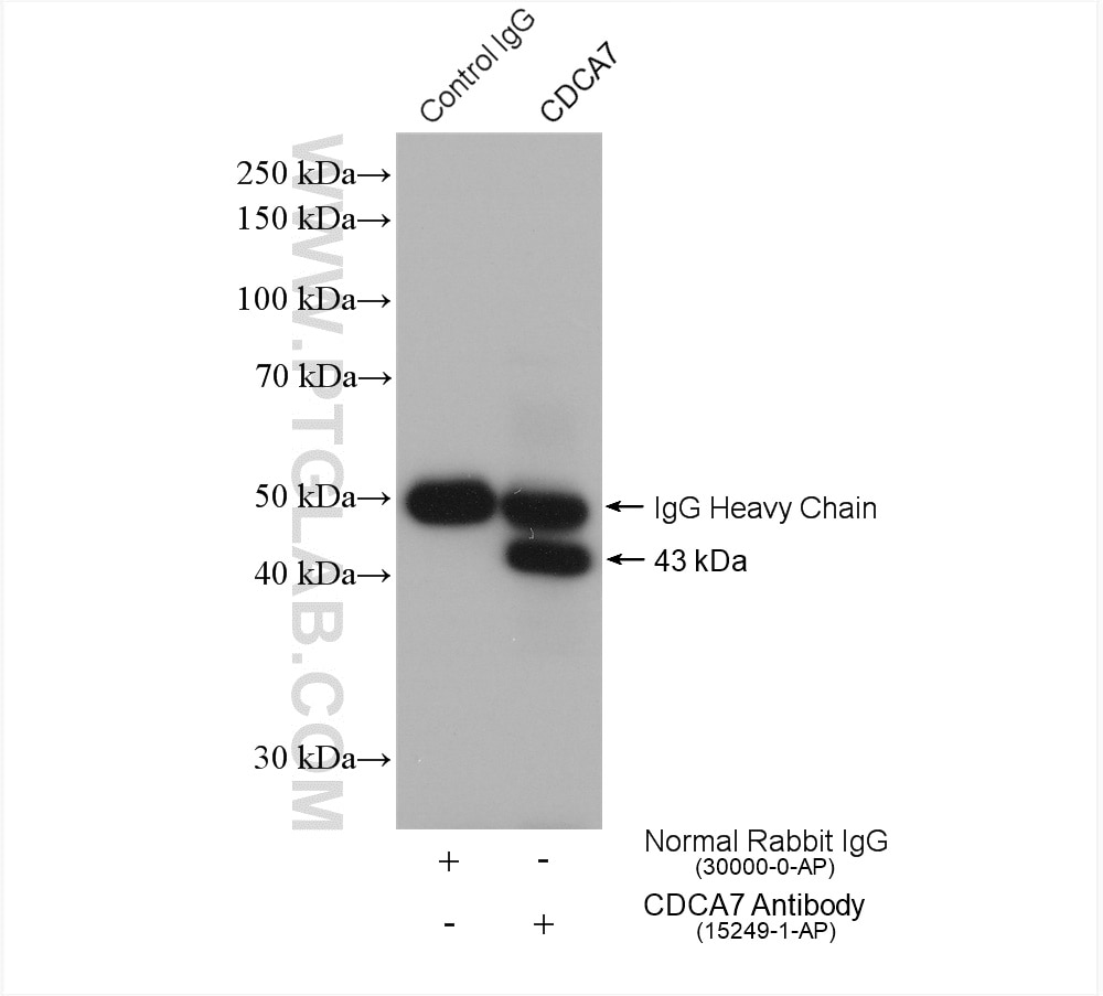
| Erfolgreiche IP | HEK-293-Zellen |
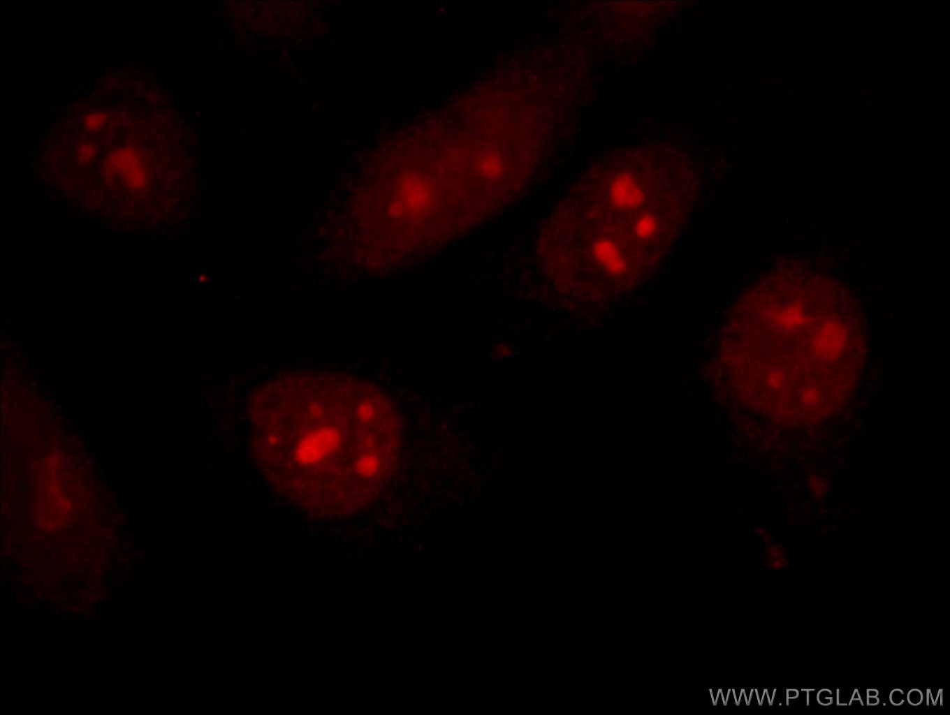
| Erfolgreiche Detektion in IF | HepG2-Zellen |
Empfohlene Verdünnung
| Anwendung | Verdünnung |
|---|---|
| Western Blot (WB) | WB : 1:1000-1:5000 |
| Immunpräzipitation (IP) | IP : 0.5-4.0 ug for 1.0-3.0 mg of total protein lysate |
| Immunfluoreszenz (IF) | IF : 1:10-1:100 |
| It is recommended that this reagent should be titrated in each testing system to obtain optimal results. | |
| Sample-dependent, check data in validation data gallery | |
Veröffentlichte Anwendungen
| WB | See 7 publications below |
| IHC | See 1 publications below |
| IF | See 1 publications below |
Produktinformation
15249-1-AP bindet in WB, IP, IHC, IF, ELISA CDCA7 und zeigt Reaktivität mit human, Maus, Ratten
| Getestete Reaktivität | human, Maus, Ratte |
| In Publikationen genannte Reaktivität | human, Maus |
| Wirt / Isotyp | Kaninchen / IgG |
| Klonalität | Polyklonal |
| Typ | Antikörper |
| Immunogen | CDCA7 fusion protein Ag4297 |
| Vollständiger Name | cell division cycle associated 7 |
| Berechnetes Molekulargewicht | 43 kDa |
| Beobachtetes Molekulargewicht | 40-43 kDa |
| GenBank-Zugangsnummer | BC027966 |
| Gene symbol | CDCA7 |
| Gene ID (NCBI) | 83879 |
| Konjugation | Unkonjugiert |
| Form | Liquid |
| Reinigungsmethode | Antigen-Affinitätsreinigung |
| Lagerungspuffer | PBS mit 0.02% Natriumazid und 50% Glycerin pH 7.3. |
| Lagerungsbedingungen | Bei -20°C lagern. Nach dem Versand ein Jahr lang stabil Aliquotieren ist bei -20oC Lagerung nicht notwendig. 20ul Größen enthalten 0,1% BSA. |
Hintergrundinformationen
CDCA7, also named as JP01, participates in MYC-mediated cell transformation and apoptosis. It plays a role as transcriptional regulator.
Protokolle
| Produktspezifische Protokolle | |
|---|---|
| WB protocol for CDCA7 antibody 15249-1-AP | Protokoll herunterladen |
| IF protocol for CDCA7 antibody 15249-1-AP | Protokoll herunterladen |
| IP protocol for CDCA7 antibody 15249-1-AP | Protokoll herunterladen |
| Standard-Protokolle | |
|---|---|
| Klicken Sie hier, um unsere Standardprotokolle anzuzeigen |
Publikationen
| Species | Application | Title |
|---|---|---|
Proc Natl Acad Sci U S A HELLS and CDCA7 comprise a bipartite nucleosome remodeling complex defective in ICF syndrome. | ||
J Exp Med Loss of ZBTB24 impairs nonhomologous end-joining and class-switch recombination in patients with ICF syndrome. | ||
Hum Mol Genet Converging disease genes in ICF syndrome: ZBTB24 controls expression of CDCA7 in mammals. | ||
Bioorg Chem Novel isatin-based hybrids as potential anti-rheumatoid arthritis drug candidates: Synthesis and biological evaluation | ||
J Biol Chem TET2-BCLAF1 transcription repression complex epigenetically regulates the expression of colorectal cancer gene Ascl2 via methylation of its promoter. | ||
Int J Biol Sci Transmissible Gastroenteritis Virus (TGEV) Infection Alters the Expression of Cellular MicroRNA Species That Affect Transcription of TGEV Gene 7. |
Rezensionen
The reviews below have been submitted by verified Proteintech customers who received an incentive forproviding their feedback.
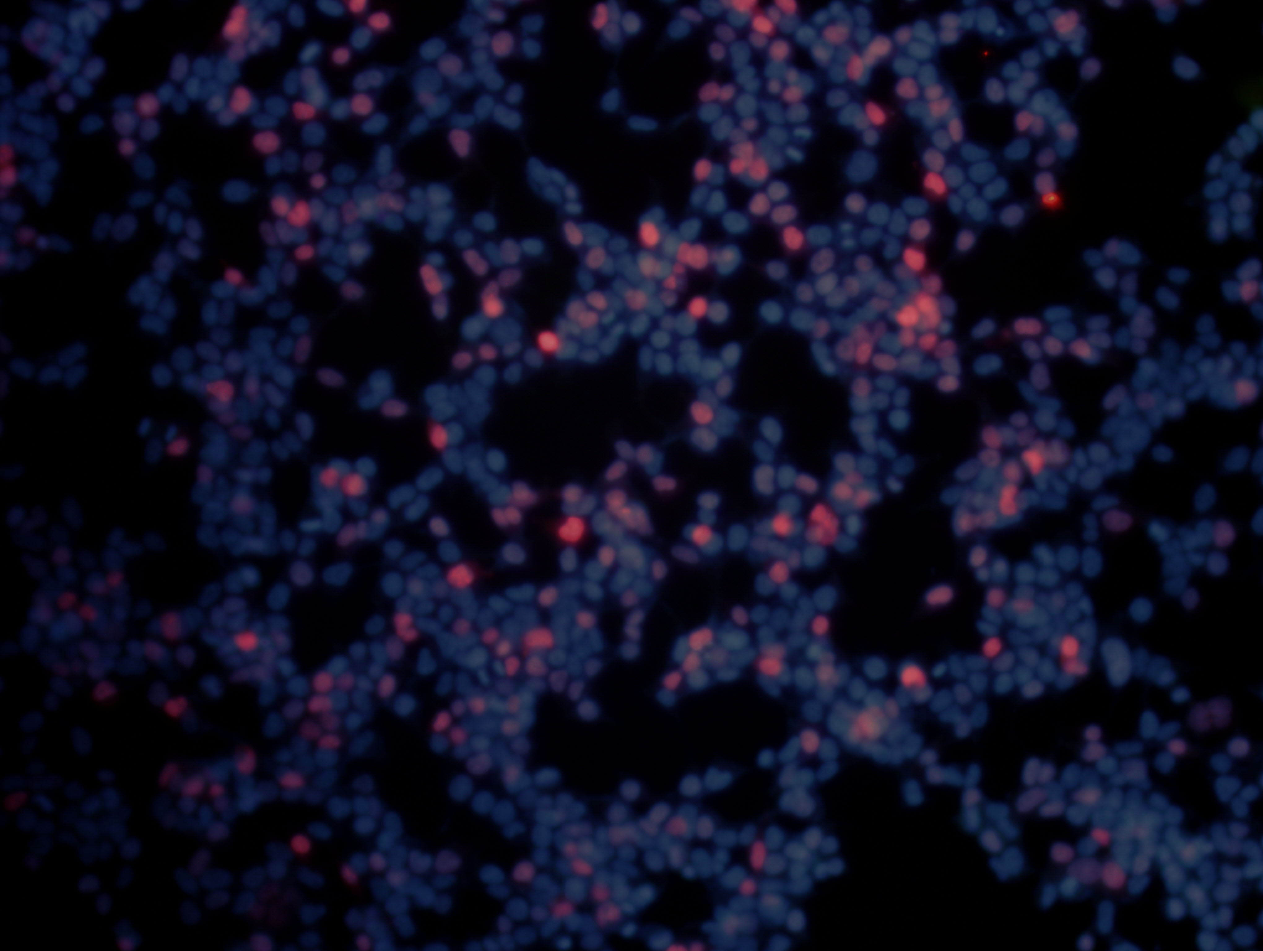
FH XIAOBING (Verified Customer) (11-05-2019) | CDCA7 antibody was used to stain the 293T cells transfected with human CDCA7 plasmid. Overexpressed CDCA7 (Red) was localized in the nucleus (DAPI was used to label the nucleus).
 |
FH Guillaume (Verified Customer) (09-08-2019) | I used cells mutated for ZBTB24 which control CDCA7 expression to test the specificity of the antibody. In WB and IP, the signal seem to be lower than the expected size in kDa. This CDCA7 antibody is not very efficient in ChIP
|
