PELP1 Polyclonal antibody
PELP1 Polyclonal Antibody for WB, IF, IHC, ELISA
Host / Isotype
Rabbit / IgG
Reactivity
Human, mouse
Applications
WB, IF, IHC, ELISA
Conjugate
Unconjugated
Cat no : 30135-1-AP
Synonyms
Validation Data Gallery
Tested Applications
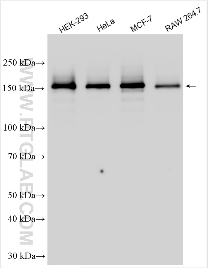
| Positive WB detected in | HEK-293 cells, HeLa cells, MCF-7 cells, RAW 264.7 cells |
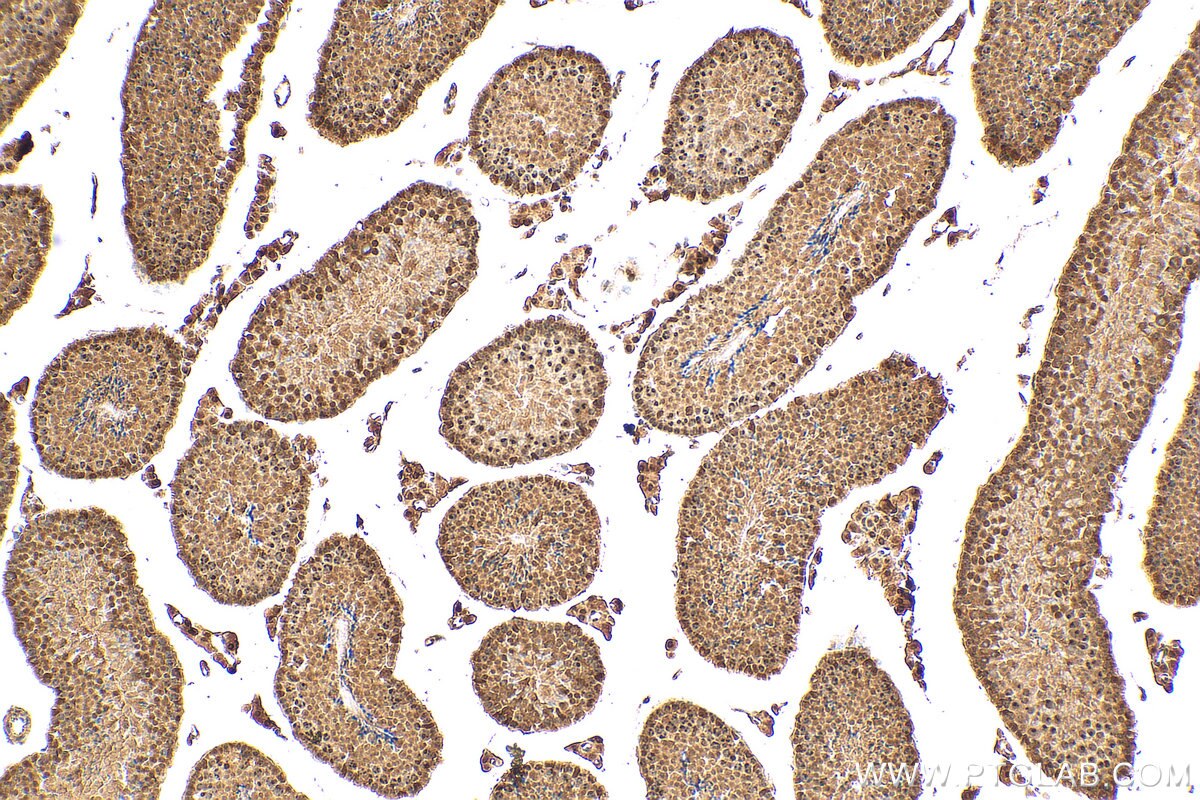
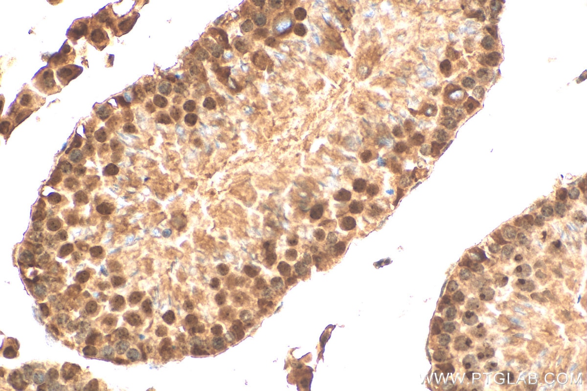
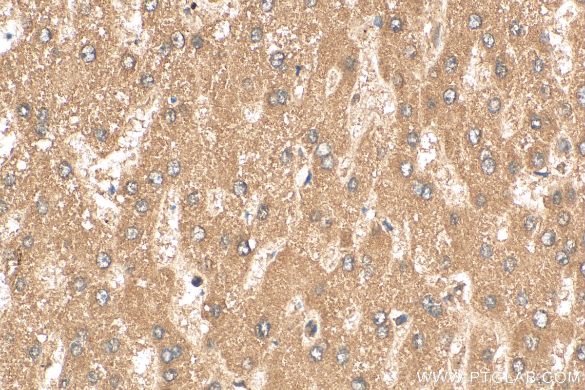
| Positive IHC detected in | mouse testis tissue, human liver tissue Note: suggested antigen retrieval with TE buffer pH 9.0; (*) Alternatively, antigen retrieval may be performed with citrate buffer pH 6.0 |
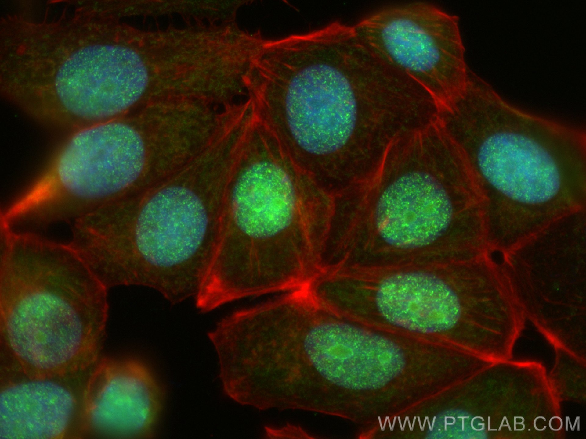
| Positive IF detected in | MCF-7 cells |
Recommended dilution
| Application | Dilution |
|---|---|
| Western Blot (WB) | WB : 1:5000-1:50000 |
| Immunohistochemistry (IHC) | IHC : 1:50-1:500 |
| Immunofluorescence (IF) | IF : 1:50-1:500 |
| It is recommended that this reagent should be titrated in each testing system to obtain optimal results. | |
| Sample-dependent, Check data in validation data gallery. | |
Product Information
30135-1-AP targets PELP1 in WB, IF, IHC, ELISA applications and shows reactivity with Human, mouse samples.
| Tested Reactivity | Human, mouse |
| Host / Isotype | Rabbit / IgG |
| Class | Polyclonal |
| Type | Antibody |
| Immunogen | PELP1 fusion protein Ag32720 |
| Full Name | proline, glutamate and leucine rich protein 1 |
| Calculated Molecular Weight | 1130 aa, 120 kDa |
| Observed Molecular Weight | 160 kDa |
| GenBank Accession Number | BC069058 |
| Gene Symbol | PELP1 |
| Gene ID (NCBI) | 27043 |
| Conjugate | Unconjugated |
| Form | Liquid |
| Purification Method | Antigen affinity purification |
| Storage Buffer | PBS with 0.02% sodium azide and 50% glycerol pH 7.3. |
| Storage Conditions | Store at -20°C. Stable for one year after shipment. Aliquoting is unnecessary for -20oC storage. 20ul sizes contain 0.1% BSA. |
Background Information
PELP1 was first identified as a 160 kDa protein in a screen for Src homology 2 (SH2) domain-binding proteins. PELP1 is overexpressed in 60-80% of breast tumors and plays important roles in both ER genomic and non-genomic signaling. In vivo, PELP1 subcellular localization is primarily nuclear in normal breast tissue, but it is localized to the cytoplasm in about 40% of invasive breast tumors. In the nucleus, PELP1 interacts with a number of transcription factors. The proto-oncogenic functions of PELP1 involve different cellular processes including epigenetic modifications leading to ER transactivation and breast cancer progression. Furthermore, PELP1 activates kinase cascades in the cytoplasm such as MAPK activation via c-Src and PI3K signaling.
Protocols
| Product Specific Protocols | |
|---|---|
| WB protocol for PELP1 antibody 30135-1-AP | Download protocol |
| IHC protocol for PELP1 antibody 30135-1-AP | Download protocol |
| IF protocol for PELP1 antibody 30135-1-AP | Download protocol |
| Standard Protocols | |
|---|---|
| Click here to view our Standard Protocols |
