- Featured Product
- KD/KO Validated
CoraLite® Plus 488-conjugated GLUT1 Monoclonal antibody
GLUT1 Monoclonal Antibody for FC (Intra)
Host / Isotype
Mouse / IgG1
Reactivity
human, mouse
Applications
FC (Intra)
Conjugate
CoraLite® Plus 488 Fluorescent Dye
CloneNo.
2A5A2
Cat no : CL488-66290
Synonyms
Validation Data Gallery
Tested Applications
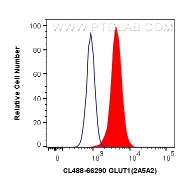
| Positive FC detected in | Jurkat cells |
Recommended dilution
| Application | Dilution |
|---|---|
| Flow Cytometry (FC) | FC : 0.40 ug per 10^6 cells in a 100 µl suspension |
| It is recommended that this reagent should be titrated in each testing system to obtain optimal results. | |
| Sample-dependent, Check data in validation data gallery. | |
Product Information
CL488-66290 targets GLUT1 in FC (Intra) applications and shows reactivity with human, mouse samples.
| Tested Reactivity | human, mouse |
| Host / Isotype | Mouse / IgG1 |
| Class | Monoclonal |
| Type | Antibody |
| Immunogen | GLUT1 fusion protein Ag17108 |
| Full Name | solute carrier family 2 (facilitated glucose transporter), member 1 |
| Calculated Molecular Weight | 492 aa, 54 kDa |
| Observed Molecular Weight | 45-55 kDa |
| GenBank Accession Number | BC121804 |
| Gene Symbol | SLC2A1 |
| Gene ID (NCBI) | 6513 |
| Conjugate | CoraLite® Plus 488 Fluorescent Dye |
| Excitation/Emission Maxima Wavelengths | 493 nm / 522 nm |
| Form | Liquid |
| Purification Method | Protein G purification |
| Storage Buffer | PBS with 50% Glycerol, 0.05% Proclin300, 0.5% BSA, pH 7.3. |
| Storage Conditions | Store at -20°C. Avoid exposure to light. Stable for one year after shipment. Aliquoting is unnecessary for -20oC storage. 20ul sizes contain 0.1% BSA. |
Background Information
Glucose transporter 1 (GLUT1), also known as solute carrier family 2, facilitated glucose transporter member 1 (SLC2A1), is a uniporter protein responsible for the transport of glucose in many cell types and across the blood-brain barrier.
What is the molecular weight of GLUT1? Is GLUT1 post-translationally modified?
There are two forms of GLUT1 transporter that differ in their molecular weight. The 45-kDa form is found in glial cells, while the 55-kDa form is present in the endothelial cells regulating glucose transport over the blood-brain and blood-tissue barriers (PMID: 9630522). N-glycosylation of asparagine at position 42 is the only known post-translation modification of GLUT1 (PMID: 3839598).
What is the subcellular localization of GLUT1?
Glucose transporters, including GLUT1, are multiple-pass integral membrane proteins. GLUT1 is present at the plasma membrane but is also a subject of recycling between plasma membrane and endosomes.
What molecules can be transported by GLUT1?
The main substrate of GLUT1 transport is glucose, but it can also transport galactose, mannose, glucosamine, and reduced ascorbate.
What is the tissue expression pattern of GLUT1?
GLUT1 is expressed by many cell types but the highest levels are observed in erythrocytes and in the central nervous system (astrocytes). GLUT1 is responsible for glucose transfer across the blood-brain and blood-tissue barriers, including placental transport.
Protocols
| Product Specific Protocols | |
|---|---|
| FC protocol for CL Plus 488 GLUT1 antibody CL488-66290 | Download protocol |
| Standard Protocols | |
|---|---|
| Click here to view our Standard Protocols |


