aequorea victoria GFP tag Monoclonal antibody
GFP tag Monoclonal Antibody for WB, IP, IF, FC, ELISA
Host / Isotype
Mouse / IgG2a
Reactivity
recombinant protein and More (5)
Applications
WB, IP, IF, FC, RIP, IHC, CoIP, ChIP, ELISA
Conjugate
Unconjugated
418
CloneNo.
1E10H7
Cat no : 66002-1-Ig
Synonyms
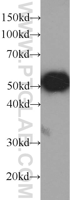
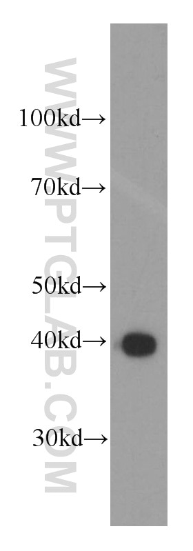
Validation Data Gallery
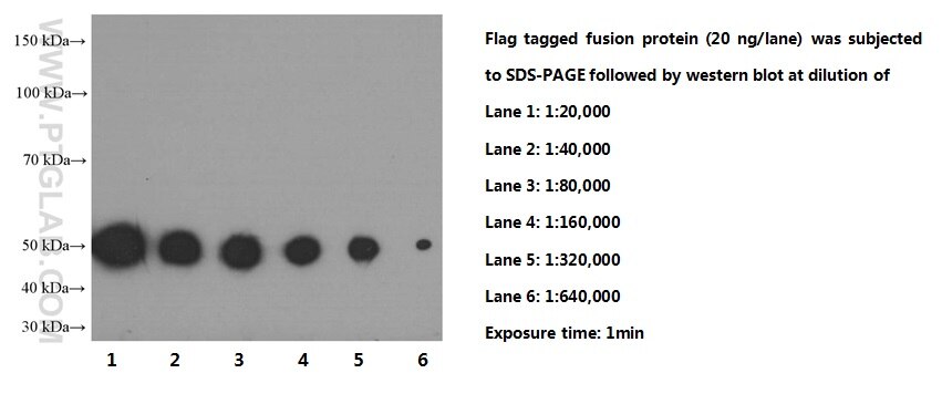
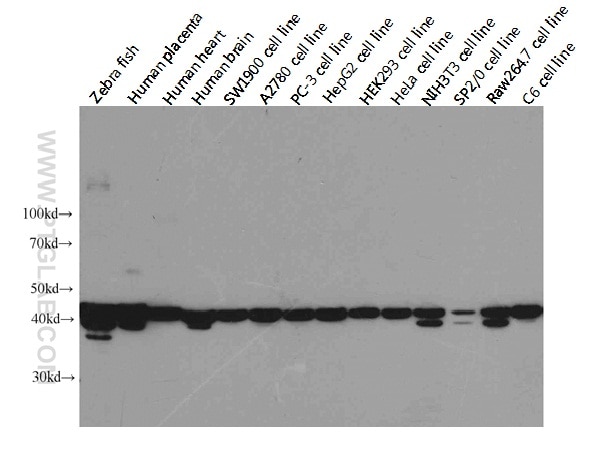
 at dilution of 1:200000 incubated at room temperature for 1.5 hours.")
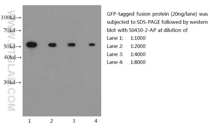
 with GFP fusion protein at varous dilutions.")
 at dilution of 1:10000 incubated at room temperature for 1.5 hours.")
 at dilution of 1:20000 incubated at room temperature for 1.5 hours.")
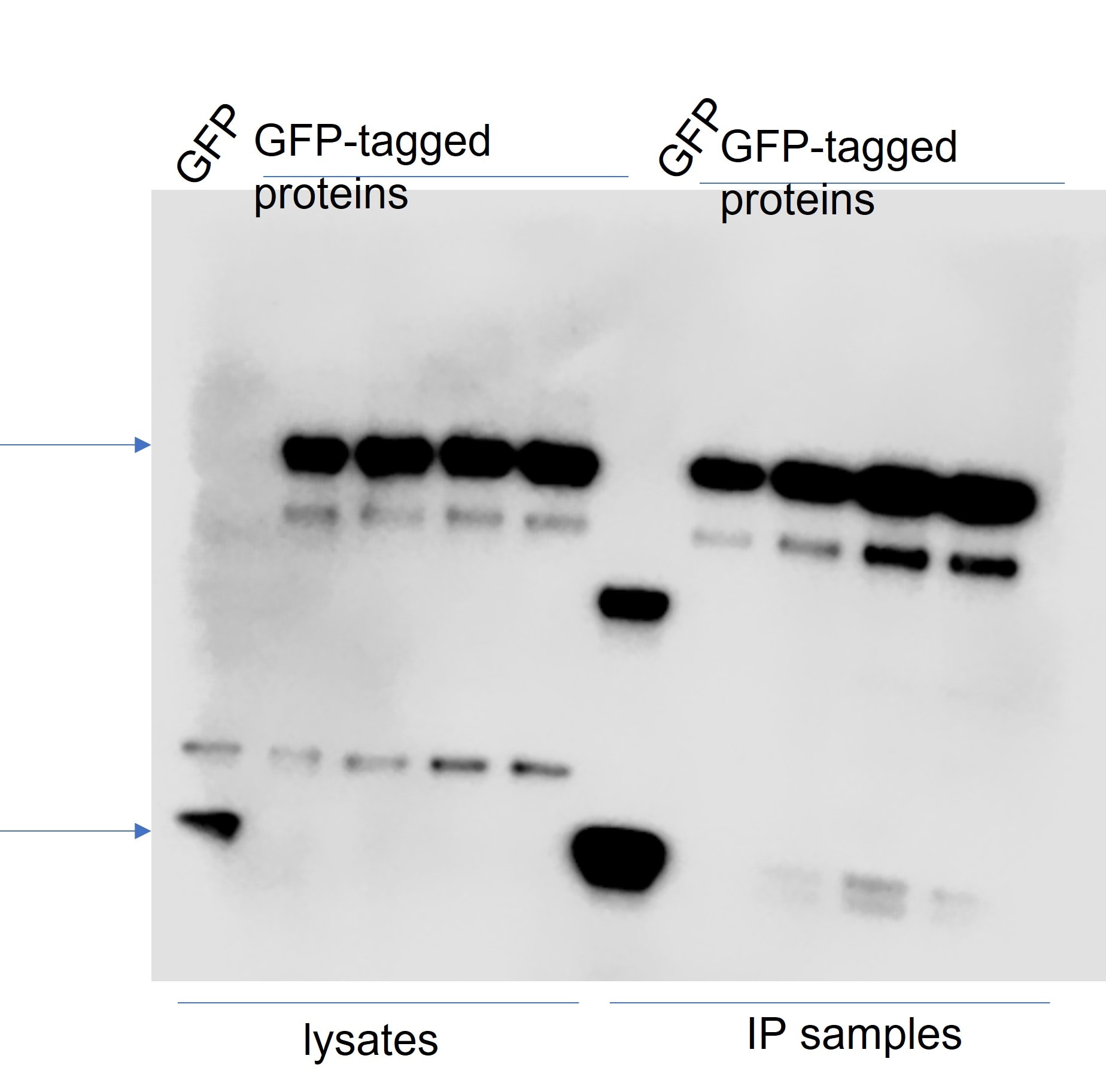
 with the vector plasmid pEGFP-N1 Transfected <a class='green' href='/productredirect?CatalogNo=HEK-293' target='_blank'>HEK-293</a> cells lysate 300ug.")
 at dilution of 1:50 and Alexa Fluor 594-conjugated AffiniPure Goat Anti-Mouse IgG (H+L).")
 and CoraLite®594-Conjugated AffiniPure Goat Anti-Mouse IgG(H+L) at dilution 1:1000 (red), and 0.2 ug Control Antibody. Cells were fixed with 90% MeOH.")
Tested Applications
| Positive WB detected in | GFP transgenic mouse brain tissue, Recombinant protein |
| Positive IP detected in | Transfected HEK-293 cells |
| Positive IF detected in | Transfected HEK-293 cells |
| Positive FC detected in | Transfected HEK-293 cells |
Recommended dilution
| Application | Dilution |
|---|---|
| Western Blot (WB) | WB : 1:20000-1:100000 |
| Immunoprecipitation (IP) | IP : 0.5-4.0 ug for 1.0-3.0 mg of total protein lysate |
| Immunofluorescence (IF) | IF : 1:20-1:200 |
| Flow Cytometry (FC) | FC : 0.20 ug per 10^6 cells in a 100 µl suspension |
| It is recommended that this reagent should be titrated in each testing system to obtain optimal results. | |
| Sample-dependent, Check data in validation data gallery. | |
Product Information
66002-1-Ig targets GFP tag in WB, IP, IF, FC, RIP, IHC, CoIP, ChIP, ELISA applications and shows reactivity with recombinant protein samples.
| Tested Reactivity | recombinant protein |
| Cited Reactivity | human, mouse, rat, zebrafish, pig |
| Host / Isotype | Mouse / IgG2a |
| Class | Monoclonal |
| Type | Antibody |
| Immunogen | GFP tag fusion protein Ag2128 |
| Full Name | GFP tag |
| Calculated Molecular Weight | 26 kDa |
| GenBank Accession Number | U73901 |
| Gene Symbol | |
| Gene ID (NCBI) | |
| RRID | AB_11182611 |
| Conjugate | Unconjugated |
| Form | Liquid |
| Purification Method | Protein A purification |
| Storage Buffer | PBS with 0.02% sodium azide and 50% glycerol pH 7.3. |
| Storage Conditions | Store at -20°C. Stable for one year after shipment. Aliquoting is unnecessary for -20oC storage. 20ul sizes contain 0.1% BSA. |
Background Information
Green fluorescence protein (GFP) is a protein composed of 238 amino acid residues (26.9kDa) derived from the jellyfish Aequorea Victoria which emits green light (emission peak at 509nm) when excited by blue light (excitation peak at 395nm). GFP, when exposed to light in the blue to ultraviolet spectrum, will show a bright green fluorescent light, making it a very useful tool in research.
What is the molecular weight of GFP?
26.9 kDa
How does GFP work?
GFP was first isolated from the jellyfish Aequorea Victoria, a source of bioluminescence, in the 1960s and in 2008 the Nobel Prize in Chemistry was awarded "for the discovery and development of the green fluorescent protein, GFP" to Osamu Shimomura and colleagues, who recognized its potential in research (PMID: 13911999). A short amino acid sequence within the protein acts as the chromophore, which absorbs UV light at 395 nm and emits green light at 509 nm.
Why is GFP a useful reporter?
When GFP was sequenced in 1992 (PMID: 1347277) it allowed scientists to express it in other organisms using transgenic techniques. It does not require cofactors to work, is non-toxic to live cells, and is relatively small, making it ideal as a "tag" for other proteins, identifiable by shining a UV light and observing the green fluorescence. The tertiary folded structure of GFP forms a chromophore at the center of a barrel shape, which protects the fluorescence-emitting amino acid chain from solvents, meaning it can function in many environments (PMID 9759496).
What are the applications for GFP?
When expressed attached to another protein, GFP can be used as a reporter gene to measure expression levels or can easily be used in fluorescence microscopy. It has been used to highlight proteins in a variety of model organisms, including bacteria, zebrafish, and mice.
Protocols
| Product Specific Protocols | |
|---|---|
| WB protocol for GFP tag antibody 66002-1-Ig | Download protocol |
| IF protocol for GFP tag antibody 66002-1-Ig | Download protocol |
| IP protocol for GFP tag antibody 66002-1-Ig | Download protocol |
| Standard Protocols | |
|---|---|
| Click here to view our Standard Protocols |
Publications
| Species | Application | Title |
|---|---|---|
Cell Discov Glc7/PP1 dephosphorylates histone H3T11 to regulate autophagy and telomere silencing in response to nutrient availability | ||
Nat Genet Pathogenic SPTBN1 variants cause an autosomal dominant neurodevelopmental syndrome. | ||
Cell Stem Cell DUX-miR-344-ZMYM2-Mediated Activation of MERVL LTRs Induces a Totipotent 2C-like State. | ||
Nat Neurosci Reversal of prolonged obesity-associated cerebrovascular dysfunction by inhibiting microglial Tak1. | ||
Nat Biomed Eng Restoration of dystrophin expression in mice by suppressing a nonsense mutation through the incorporation of unnatural amino acids. |
Reviews
The reviews below have been submitted by verified Proteintech customers who received an incentive forproviding their feedback.
FH Amy (Verified Customer) (02-11-2023) | Detected EGFP-tagged constructs expressed in HEK293T cells at similar strength to other antibodies however with slightly more background.
 |
FH Tom (Verified Customer) (02-03-2023) | Works great. No problem. Love proteintech.
|
FH Tatyana (Verified Customer) (01-09-2023) | Good signal, antibody can be reused. Overexpressed GFP-tagged proteins (and GFP alone) were pulled down using GFP-Trap beads and WB was done on lysates and IP samples. Antibody was diluted in 4% milk (and could be reused several times).
 |
FH Sheila (Verified Customer) (07-22-2022) | It works very well
|
FH Juliana (Verified Customer) (01-28-2022) | works great for Western blot!
|
FH Prasanna (Verified Customer) (01-04-2021) | Of the 3 antibodies tested for GFP this one gave the cleanest and strongest signal by western blot. When I saw it on sale, I bought 8!
|
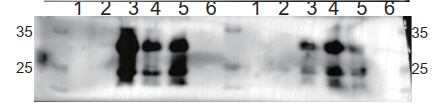
FH Lana (Verified Customer) (12-22-2020) | SDS-PAGE: 15 ug/ul RIPA protein lysate, 4-12% Bis-Tris gradient gel.Transfer: Immobilon-FL transfer membranes (Millipore) for 2h at 80V, 4C.Blocking: SEA Block Blocking Buffer 1h, room T.Primary Ab: O/N incubation at 4C, 1:5000.Secondary Ab: IRDye 800CW Goat anti-Mouse, 1:15000.Lines of WB image: 1 – protein ladder, 2 – HEK293 whole cell lysate, negative transfection, 3 – whole cell lysate of cells transfected with eGFP.
 |
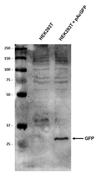
FH Thomas (Verified Customer) (11-19-2020) | HEK293T and HEK293T stably transfected with pAcGFP plasmid. 10ug total protein loaded per well. Membrane blocked 1 hour in 5% BSA prior to anti-GFP (1:2000) o/n at 4 degrees. Goat anti-mouse HRP secondary (1:10,000) used.
 |
FH Jane (Verified Customer) (03-02-2020) | Adenovirus-GFP infected cardiomyocytes stained with GFP antibody, signal is strong and effective
|
FH LUNFENG (Verified Customer) (01-27-2020) | GOOD
|
FH Jie (Verified Customer) (01-27-2020) | Worked for western blot with GFP-LC3 transfected cardiomyocytes
|
FH Paul (Verified Customer) (01-15-2020) | Works well for Westerns.
|
FH Laura (Verified Customer) (01-15-2020) | Good antibody for Western Blot.
|
FH Aamir (Verified Customer) (01-08-2020) | Worked well for WB
|
FH Benjamin (Verified Customer) (01-07-2020) | Easily detects recombinant GFP protein via western blot with very little background.
|
FH Jing (Verified Customer) (01-03-2020) | Used this to detect tranduce efficiency of the AAV-GFP virus. with 5% non-fat milk, there are two bands around 20-30kd, not sure which one is correct. And the antibody is relative weak, has to use 1:500 dilution.
|
FH Shan (Verified Customer) (12-25-2019) | The GFP antibody showed great sensitivity for WB and it was easily detecable. But it was insteresting that when the SDS PAGE gel separating gel concentration reeached to 12%, you can see two bands at ~30kDa and ~20kDa.
 |
FH Jason (Verified Customer) (11-04-2019) | This is a good antibody for detecting GFP tag on Western blot, using 1:1000 dilution. The price is unbeatable, worth each penny.
|
FH Hend (Verified Customer) (10-14-2019) | antibody used in western blot against egfp labelled protien and clear band was detected.
|