How To Optimize Your ELISA Experiments
Technical tips for better ELISA results
|
Matched antibody pair in ELISA kit ELISA blocking and washing steps |
Introduction to ELISA
The enzyme-linked immunosorbent assay (ELISA) is one of the most sensitive and reproducible technologies available. These assays are rapid, simple to perform, and easily automated. As with any assay, the reproducibility and reliability of ELISAs depend upon proper technique and attention to detail.
The ELISA technique is divided into:
-
Direct ELISA, where the antigen is immobilized on the ELISA plate, and the primary antibody carries the label (Figure 1 A)
-
Indirect ELISA, where the antigen is immobilized on the ELISA plate, and the secondary antibody carries the label (Figure 1 B)
-
Sandwich ELISA, where two primary antibodies (for capture and detection) embed the antigen, forming a "sandwich" and then the complex is recognized by a secondary labelled antibody (Figure 1 C).
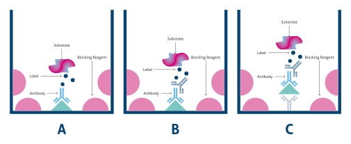
Figure 1. Types of ELISA formats: A) direct, B) indirect and C) Sandwich ELISA.
Matched antibody pair in ELISA kit
Matched pair refers to sets of antibodies that are known to recognize different epitopes on the same protein antigen, so they can be used together for the capture and detection of a single antigen in a sandwich ELISA. The antibodies used in ELISA assays can be monoclonal, polyclonal, or a combination of both.
Monoclonal antibodies can be used for all antibody-containing steps in all types of ELISAs. They are commonly used in sets as matched pairs in sandwich ELISAs but can be used for capture or detection in conjunction with a polyclonal antibody to enhance signal or to provide a greater chance of capturing an antigen from a complex solution.
Due to the heterogeneity of antibodies present in polyclonal antibody solutions and the wide representation of epitopes present, polyclonal antibodies can be powerful tools for the detection of an antigen, often yielding higher signal levels than monoclonal antibodies. However, polyclonal antibodies are more likely to share one or more epitopes with closely related proteins, resulting in higher non-specific signal. One method of reducing this problem is to use affinity purified or cross-absorbed polyclonal antibodies. To increase assay sensitivity, the detection method for an ELISA can be switched from direct to indirect detection using a polyclonal antibody.
ELISA samples
A variety of samples can be tested in an ELISA, and the choice of assay conditions will depend upon the complexity of the sample and the expected amount of antigen present.
It is important to test all samples in duplicate or triplicate in conjunction with a known standard to ensure the accuracy of results and for quantitation.
It is better to test several dilutions of a sample to make sure the final results fall within the linear portion of the standard curve. Highly concentrated samples can underestimate concentration while highly diluted samples can overestimate concentrations.
ELISA blocking and washing steps
Blocking is often necessary to prevent non-specific binding of detection antibodies to the multi-well plate surface itself. When a plate is fully blocked, assay sensitivity will be enhanced since non-specific signal will be reduced.
A thorough washing procedure is essential for obtaining reliable ELISA results. It is important to completely aspirate liquid from all wells by gently lowering an aspiration tip into the bottom. Avoid scratching the inside of the well. When washing is complete, it is recommended to invert the plate and dry it on absorbent tissue.
How to optimize your ELISA experiment
Although each component is described separately, in many cases it is possible to optimize two components simultaneously by performing a checkerboard titration.
1) Capture Antibody Concentration
- Prepare different concentrations of the capture antibody in coating buffer.
2) The Blocking Buffer
- Prepare different blocking solutions. If the blocking solution is not preformulated (i.e., it is a single protein, such as BSA), try different concentrations of the protein.
3) The Standard Diluent
-
Try to match the standard diluent as closely as possible to the matrix of the sample.
-
If the matrix itself cannot be exactly duplicated then test different standard diluent solutions and check the standard curve and linearity of dilution for the sample. It may be necessary to choose a different diluent.
- If the sample has poor linearity, there may be an imbalance between the sample matrix and the standard diluent. In such cases spike-and-recovery or linearity-of-dilution experiments should be performed.
4) Sample Concentration
- Prepare different concentrations of the sample, keeping in mind the detection limit of the substrate. To confirm that the biological sample matrix is not masking or enhancing the signal, spike-and-recovery and linearity-of dilution experiments should be performed.
5) The Detection Antibody Concentration
- Prepare different concentrations of the detection antibody.
6) The Enzyme Conjugate Concentration
- Prepare different concentrations of the enzyme conjugate according to the ELISA kit range described for the substrate.
7) Signal Detection
-
Select substrate(s) based on the amount of the antigen in the sample and ability to detect it with a plate reader.
-
If the antigen can clearly be detected then the substrate is appropriate.
-
If the antigen is below the threshold for detection then select a more sensitive substrate.
Common ELISA issues
1) Weak or no signal in ELISA
-
It is recommended that all reagents are at room temperature for 15–20 minutes before starting the assay.
-
Incorrect storage of components. Double-check storage conditions on the kit label. Most kits need to be stored at 2–8oC.
-
Reagents added/prepared incorrectly. Check the protocol, ensure reagents were added in the proper order, and prepared to correct dilution.
-
Overexposure of fluorescent reagents. Intense light can cause photo-bleaching by decomposition of the fluorophore. Protection of the fluorophore from light is essential for effective signal generation at the end of the assay.
-
Components require further optimization. One of the components of the assay may be at a limiting concentration, resulting in lower overall signal.
-
Degradation of reagents. One of the reagents may have degraded or been contaminated, in which case it should be replaced.
-
Antibodies are an inefficient pair or lack sufficient affinity towards the target. The antibodies used may not bind effectively to the antigen or may not work in combination with each other. In such cases, alternative antibodies must be tested.
-
Detection system requires further optimization. The detection system (substrate) may not be sensitive enough to give the signal, or the standard curve may not be appropriate for the sample. It might be necessary to concentrate the sample or switch to a more sensitive substrate.
2) High background
-
Insufficient washing or blocking. High background signal is commonly the result of either insufficient washing or blocking, sample components or antibodies cross-reacting with the blocking buffer, or the use of too much enzyme conjugate.
-
It is important to balance the amount of enzyme giving specific signal versus that giving background signal. The most effective way to control this is by optimizing the enzyme conjugate, antibodies, and blocking solution.
3) Poor standard curve linearity
-
If the standard curve has a poor linearity, then samples must fall within a tight concentration range to be deemed accurate.
-
If the curve has good linearity but poor variation between replicates (i.e., standard error), there might be a technical problem such as inconsistent pipetting between samples or individual users.
All samples and standards should be measured at least in duplicate or triplicate to mitigate this issue.
4) Excessively high signal in ELISA
-
Insufficient washing. To avoid this issue, use the appropriate washing procedure, e.g., at the end of each washing step, invert the plate on absorbent tissue and allow to completely drain, tapping forcefully if necessary to remove any residual fluid.
-
Incubation time is too long. Excessive incubation time is also a reason for overly high signal in ELISA; be sure to follow recommended incubation times.
-
Contamination. Avoid this issue using plate sealers, and use a fresh sealer each time the plate is opened. This will prevent wells from contaminating each other.


Support
Newsletter Signup
Stay up-to-date with our latest news and events. New to Proteintech? Get 10% off your first order when you sign up.