Filter:
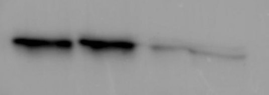
 with sh-Control and sh-CNN3 transfected HEK-293 cells.")
 lysates prepared with RIPA buffer, 20 μg of protein loaded. 11509-1-AP incubated at 1:100 at 4°C overnight in 5% milk in TBST. Ponceau stained transfers shown for each blot.
These data were performed and provided by YCharOS in partnership with Proteintech. YCharOS is an open science company with a mission to characterise all commercial antibodies to improve scientific reproducibility and transparency.")
 at dilution of 1:500 incubated at room temperature for 1.5 hours.")
 at dilution of 1:3000 incubated at room temperature for 1.5 hours.")
 at dilution of 1:500 incubated at room temperature for 1.5 hours.")

 with HEK-293 cells lysate 960 ug.")
 at dilution of 1:200 (under 10x lens). Heat mediated antigen retrieval with Tris-EDTA buffer (pH 9.0).")
 at dilution of 1:200 (under 40x lens). Heat mediated antigen retrieval with Tris-EDTA buffer (pH 9.0).")
 at dilution of 1:4000 (under 20x lens). Heat mediated antigen retrieval with Tris-EDTA buffer (pH 9.0).")
 at dilution of 1:50 (under 10x lens).")
 at dilution of 1:50 (under 40x lens).")
 at dilution of 1:50 (under 10x lens).")
 at dilution of 1:50 (under 40x lens).")
 fixed HeLa cells using CNN3 antibody (11509-1-AP) at dilution of 1:400 and Multi-rAb CoraLite ® Plus 488-Goat Anti-Rabbit Recombinant Secondary Antibody (H+L) (RGAR002).")
Tested Applications
| Positive WB detected in | HCT 116 cells, HAP1 cells, human kidney tissue, human skeletal muscle tissue, HEK-293 cells, HepG2 cells, NIH/3T3 cells |
| Positive IP detected in | HEK-293 cells, U-87MG cells |
| Positive IHC detected in | human prostate cancer tissue, human hysteromyoma tissue, human skin tissue, human stomach cancer tissue Note: suggested antigen retrieval with TE buffer pH 9.0; (*) Alternatively, antigen retrieval may be performed with citrate buffer pH 6.0 |
| Positive IF/ICC detected in | HeLa cells |
Recommended dilution
| Application | Dilution |
|---|---|
| Western Blot (WB) | WB : 1:1000-1:6000 |
| Immunoprecipitation (IP) | IP : 0.5-4.0 ug for 1.0-3.0 mg of total protein lysate |
| Immunohistochemistry (IHC) | IHC : 1:50-1:500 |
| Immunofluorescence (IF)/ICC | IF/ICC : 1:200-1:800 |
| It is recommended that this reagent should be titrated in each testing system to obtain optimal results. | |
| Sample-dependent, Check data in validation data gallery. | |
Published Applications
| KD/KO | See 1 publications below |
| WB | See 2 publications below |
| IHC | See 1 publications below |
| IF | See 1 publications below |
Product Information
11509-1-AP targets CNN3 in WB, IHC, IF/ICC, IP, ELISA applications and shows reactivity with human, mouse, rat samples.
| Tested Reactivity | human, mouse, rat |
| Cited Reactivity | human, mouse |
| Host / Isotype | Rabbit / IgG |
| Class | Polyclonal |
| Type | Antibody |
| Immunogen | CNN3 fusion protein Ag2068 Predict reactive species |
| Full Name | calponin 3, acidic |
| Calculated Molecular Weight | 329 aa, 36 kDa |
| Observed Molecular Weight | 36 kDa |
| GenBank Accession Number | BC025372 |
| Gene Symbol | CNN3 |
| Gene ID (NCBI) | 1266 |
| RRID | AB_2082140 |
| Conjugate | Unconjugated |
| Form | Liquid |
| Purification Method | Antigen affinity purification |
| UNIPROT ID | Q15417 |
| Storage Buffer | PBS with 0.02% sodium azide and 50% glycerol , pH 7.3 |
| Storage Conditions | Store at -20°C. Stable for one year after shipment. Aliquoting is unnecessary for -20oC storage. 20ul sizes contain 0.1% BSA. |
Protocols
| Product Specific Protocols | |
|---|---|
| WB protocol for CNN3 antibody 11509-1-AP | Download protocol |
| IHC protocol for CNN3 antibody 11509-1-AP | Download protocol |
| IF protocol for CNN3 antibody 11509-1-AP | Download protocol |
| IP protocol for CNN3 antibody 11509-1-AP | Download protocol |
| Standard Protocols | |
|---|---|
| Click here to view our Standard Protocols |
Publications
| Species | Application | Title |
|---|---|---|
Cell Rep Spatiotemporal dynamics of inner ear sensory and non-sensory cells revealed by single-cell transcriptomics. | ||
J Proteome Res Comparative Proteomics Reveals Prolonged Corneal Preservation Impaired Ocular Surface Immunity Accompanied by Fibrosis in Human Stroma | ||
Reviews
The reviews below have been submitted by verified Proteintech customers who received an incentive for providing their feedback.
FH Elena (Verified Customer) (01-31-2025) | Great antibody. CNN3 antibody was used for Western blot for two different breast cancer cell lines. Very good signal in just 1 min of exposure with low background.
 |


